• Skip to primary navigation
  • Skip to main content
  • Skip to primary sidebar
  • Skip to footer

Johns Hopkins Vasculitis Center

Show Search
Hide Search
  • About Our Center
    • Appointments and Directions
    • Meet Our Team
    • Support Our Center
  • What is Vasculitis?
    • Types of Vasculitis
    • Causes of Vasculitis
    • Symptoms of Vasculitis
    • Diagnosing Vasculitis
  • Vasculitis Treatments
    • Prednisone
    • Avacopan (Tavneos®)
    • Apremilast (Otezla®)
    • Azathioprine
    • Colchicine
    • Cyclophosphamide (Cytoxan)
    • Dapsone
    • Supplemental Immunoglobulin (IVIG/SCIG)
    • Leflunomide
    • Mepolizumab (Nucala®)
    • Methotrexate (MTX)
    • Mycophenolate
    • Rituximab
    • Sarilumab (Kevzara®)
    • TNF Inhibitors
    • Tocilizumab (Actemra®)
  • Vasculitis Research
  • Resources
Home / Search for "search.html/whatis/polyangiitis.html/page/cryoglobulinemia.html"

Search Results for: search.html/whatis/polyangiitis.html/page/cryoglobulinemia.html

Glossary of Vasculitis Terms

A

Active disease: Disease state characterized by active, ongoing inflammation due to vasculitis. In this state, vasculitis causes damage to organs. Treatment is initiated to induce disease remission.

Alkylating agents: A group of drugs originally used to treat cancer, now used (in lower doses) to treat some forms of severe vasculitis.

ANCA: Anti-neutrophil cytoplasmic antibodies. The abbreviation is pronounced just like the last name of the singer, Paul Anka. These antibodies are found in patients with some forms of vasculitis, particularly granulomatosis with polyangiitis, microscopic polyangiitis, and the Churg-Strauss syndrome.

Anemia: A low hematocrit (roughly corresponding to a low red blood cell count).

Anesthesic: Means literally “without feeling”. A drug administered for medical or surgical purposes, that induces partial or total loss of sensation. An anesthesic may be topical, local, regional, or general, depending on the method of administration and area of the body affected.

Aneurysms: Weakening of a blood vessel wall by inflammation. Sometimes leads to rupture of the vessel.

Angiogram: An X-ray representation of blood vessels made after the injection of a radio-opaque dye. Used to visualize the inner layer of blood vessels and to determine the location and degree of narrowing or dilation.

Antibodies: A Y-shaped protein on the surface of B cells that is secreted into the blood in response to a challenge to the immune system, such as a bacterium, virus, parasite, or transplanted organ. Antibodies neutralize foreign proteins by binding specifically to them. Antibodies are also known as immunoglobulins.

Aphthous ulcers: Ulcerations on the mucous membranes of the mouth or genitals, often caused by an infection or trauma. Aphthous ulcers also occur in Behcet’s disease and connective tissue diseases such as Lupus.

Arthralgias: Aches or pains in joints, without obvious swelling, warmth, or redness.

Arthritis: Inflammation of a joint, usually accompanied by pain, swelling, and stiffness, and resulting from infection, trauma, degenerative changes, metabolic disturbances, or other causes. Arthritis occurs in various forms, such as the arthritis associated with infections, osteoarthritis, or rheumatoid arthritis. Many forms of vasculitis can also be associated with arthritis.

Aseptic: Using sterile techniques or methods to protect against infection by microbes.

Asymmetrical: Having no balance or symmetry. Different on the left side of the body as compared to the right. “Asymmetric weakness of the left lower extremity” means that the left leg is weak but the right is not.

Autoimmune: Relating to an immune response by the body against one of its own tissues; for example, its own cells, molecules, or organs. Autoimmune diseases often involve the formation of antibodies directed against specific body tissues, such as parts of the kidney or blood vessel walls.

B

Beading: The appearance of a blood vessel as of a string of beads, with alternating areas of narrowing and dilation.

Biopsy: Removal of a piece of a tissue or organ through either surgery or sampling with a needle to determine the existence or cause of a disease.

C

Capillaries: The smallest blood vessel in the body. Capillaries connect arterioles (small arteries) with venules (small veins). Capillaries form an intricate network throughout the body for the interchange of various substances, such as oxygen and carbon dioxide, between blood and tissue cells.

Casts: A cluster of cells or proteins formed in the kidney and excreted in the urine. Red blood cell casts usually indicate the presence of active vasculitis within the kidney. Kidney vasculitis is also known as glomerulonephritis.

CAT Scan (CT scan): A Computerized Axial Tomography scan is an x-ray tube that rotates in a circle around the patient, making many pictures as it rotates. The multiple x-ray pictures are reconstructed by a computer in cross-sectional images, permitting doctors to examine “slices” through different organs.

Catheterization: The insertion of a small plastic tube into a blood vessel, for the purpose of infusing fluid or radio-opaque dye (as in angiography), or for the purpose of sampling blood.

Cavity: A hollow area within the body.

Chlorambucil: A medication used to depress lymphocytic production and maturation. The brand name for this medication is Leukeran.

Claudication: A symptom caused by lack of blood flow to the muscles caused by narrowing of the arteries. The symptom of claudication usually occurs in the calf or in an arm, and is an aching pain that resolves with rest.

Conjunctivitis: Inflammation of the most superficial layer of the eye, characterized by redness and often accompanied by a discharge. Conjunctivitis can be caused by certain infections, but is also associated with some types of vasculitis.

Constitutional: Symptoms Symptoms that relate to the entire body as a whole, rather than to an individual organ. Constitutional symptoms include fatigue, malaise, and weight loss.

Crescent: A shape resembling the curved shape of the moon in its first or last quarters. With regard to vasculitis, a crescent is a pathological finding in the kidney that is observed under the microscope. Crescents are caused by damage to glomeruli, the individual blood-filtering units within the kidney.

Cutaneous: Relating to, existing on, or affecting the skin.

Cyclophospamide: An alkylating agent used in combination with corticosteroids (such as prednisone) to treat serve cases of vasculitis.

Cystoscopy: The inspection of the interior of the bladder using a lighted tubular endoscope, inserted through the urethra. The major reason for performing cystoscopy in patients with vasculitis is to screen for bladder injury caused by cyclophosphamide.

Cytoplasmic: The protoplasm outside the nucleus of a cell.

D

Dermis: The layer of the skin located below the epidermis, containing nerve endings, sweat and sebaceous glands, and blood and lymph vessels. Small and medium-vessel forms of vasculitis affect the dermis and sometimes the layer just below the dermis; the subcutaneous fat.

Dialysis: A pathological deficiency in the oxygen-carrying component of the blood, measured in unit volume concentrations of hemoglobin, red blood cell volume, or red blood cell number.

E

Eosinophils: A type of white blood cell containing cytoplasmic granules that are stained easily by eosin or other acid dyes.

Epidermis: The protective outer layer of the skin.

ESR: Erythrocyte Sedimentation Rate is the rate at which red blood cells settle out in a tube of blood under standardized conditions; a high rate usually indicates the presence of inflammation.

Etiology: The cause or origin of a disease.

G

Gangrene: Death and decay of body tissue, often occurring in a limb, caused by insufficient blood supply and usually following injury or disease.

Glomerulonephritis: Inflammation in the kidney, characterized often by decreased production of urine and by the presence of blood and protein in the urine.

Glomerulus: A tuft of capillaries within the kidney that filters blood in order to form urine. Normally, each kidney has approximately 1 million glomeruli.

H

Hematuria: The presence of blood in the urine.

Hyperpigmented: An excess of pigment (color) in a tissue. For example, in some patients, parts of the skin affected by vasculitis become hyperpigmented after the vasculitis has resolved.

I

IgA: Immunoglobulin A. A specific subcategory of antibodies (which all individuals have). For reasons that are not understood, IgA deposits within the blood vessels of the skin, joints, kidney, and GI tract in Henoch-Schonlein purpura, leading to vasculitis.

Incident/incidence: The number of new cases of a disease in a population over a period of time. Physicians often refer to the “annual incidence” of a given disease; that is, the number of new cases occurring each year.

Infarction: Localized necrosis resulting from obstruction of the blood supply. Myocardial infarction is another name for a heart attack.

Infection: Invasion by and multiplication of pathogenic microorganisms in part of the body, or in the body’s bloodstream.

Infiltrates: A collection of inflammatory cells within a body tissue; for example, in the lung. Pulmonary infiltrates are visible on chest x-rays in pneumonia and in some forms of vasculitis.

Inflammation: A localized protective response of the body tissues to injury, irritation, or infection; characterized by pain, swelling, redness, and heat. Vasculitis is inflammation within the blood vessels.

Intravenous: A drug, nutrient solution, or other substance administered into a vein.

Invasive: Relating to a medical procedure in which a part of the body is entered, as by puncture or incision. Invasive procedures such as tissue biopsies or angiograms are sometimes necessary to diagnose vasculitis.

Ischemia: A decrease in the blood supply to an organ, tissue, or body part caused by constriction or obstruction of the blood vessels.

L

Lesion: A wound or injury. A localized pathological change in a bodily organ or tissue. An infected or diseased patch of skin.

Localized: Restricted or limited to a specific body part or region: localized pain and numbness.

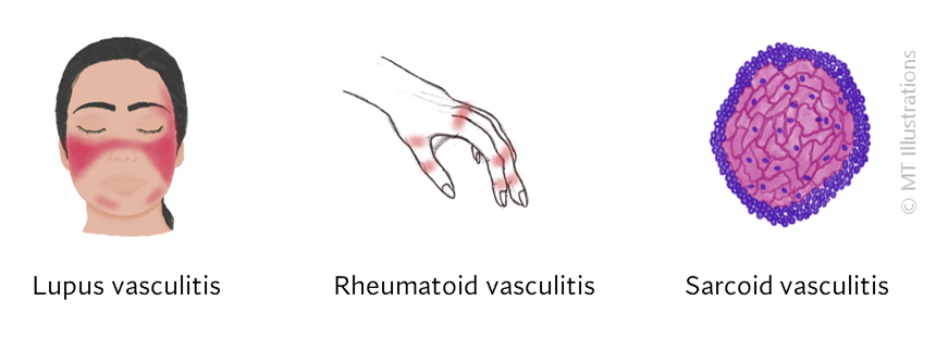
Lupus: A systemic disease that results from an autoimmune mechanism. Individuals with lupus produce antibodies to their own body tissues. The resultant inflammation can cause kidney damage, arthritis, pericarditis, and, sometimes, vasculitis.

M

Meninges: The three membranes that cover the brain and spinal cord. The three layers are called the arachnoid (the “spider-like” innermost layer), the pia mater (the middle layer), and the dura mater (the “tough mother” outermost layer).

Mesenteric: Relating to folds of the peritoneum (the abdominal cavity) that connect the intestines to the abdominal wall, especially such a fold that envelops the small intestine.

Mimicker: A disease process that imitates or simulates another. For example, the lung lesions of granulomatosis with polyangiitis may be mimickers of tuberculosis.

MRI Magnetic Resonance Imaging: Another fancy x-ray, similar to a CT scan. MRI scans also provide cross-sectional images of body organs. Because MRI technology involves the use of a large magnet, people with pacemakers, metallic aneurysm clips, and other metallic inserts are not eligible to have these studies.

Myeloperoxidase Abbreviated MPO: An enzyme found in many tissues and cells throughout the body. For reasons that are unknown, many patients with granulomatosis with polyangiitis and microscopic polyangiitis make antibodies to this protein.

N

Nasal septum: A thin wall dividing the nasal cavity into two halves.

Neuropathy: A disease or abnormality of peripheral nerves, the nerves that mediate sensation and movement in the arms, legs, and other body parts.

O

Occlusion: An obstruction or a closure of a blood vessel.

Opportunistic infections: A pathological deficiency in the oxygen-carrying component of the blood, measured in unit volume concentrations of hemoglobin, red blood cell volume, or red blood cell number.

Otitis media: Inflammation of the middle ear (the space just behind the eardrum). This may occur because of an infection or as a result of some forms of vasculitis, such as granulomatosis with polyangiitis.

P

Palpable: Perceptible to touch; capable of being palpated.

Pathologist: A physician who examines tissue biopsies for the purpose of determining the precise cause of disease.

Perforation: A hole in an organ, such as the gastrointestinal tract.

Perinuclear: Of or pertaining to a nucleus; situated around a nucleus. In some forms of vasculitis (e.g., microscopic polyangiitis), anti-neutrophil cytoplasmic antibodies (ANCA) cause perinuclear staining on immunofluorescence tests.

Pneumonia: An acute or chronic disease marked by inflammation of the lungs and caused by viruses, bacteria, or other microorganisms. Forms of vasculitis that involve the lung are often misdiagnosed as pneumonia. Pneumonia is also a type of opportunistic infection that may occur in vasculitis patients under treatment.

Prevalence: The total number of cases of a disease present within a given population at a particular time. The prevalence of giant cell arteritis in the United States, for example is estimated to be 160,000.

Proteinuria: The presence of excessive amounts of protein in the urine. Proteinuria is usually caused by damage to the kidneys.

Purpura: A condition characterized by small amounts of bleeding into the skin and mucous membranes that result in the appearance of purplish spots or patches.

R

Raynaud’s phenomenon: Spasm of the arteries to the fingers and/or toes, resulting in blanching or pain.

Remission: When vasculitis has been treated to the point that there are no signs or symptoms of ongoing active inflammation. In this state, patient may experience symptoms related to damage from prior disease activity, but no active inflammation is present.

Retinal: The innermost layer of the eye. Serves as the eye’s camera, transmitting images to the brain via the optic nerve.

Rheumatologist: A physician who specializes in the diagnosis and treatment of diseases in which the immune system has gone awry, leading to inflammation in a variety of organs.

S

Sclera: The white part of the eye. “Scleritis” is a type of inflammation that occurs in the sclera in some forms of vasculitis.

Stenosis: A constriction or narrowing of a blood vessel.

Steroid-sparing drug: An immunosuppressive drug that has the benefits of prednisone but does not cause as many side effects.

Subcutaneous: Underneath the skin. Some medications, for example, are injected under the skin.

Systemic: Relating to a system or relating to, or affecting the entire body or an entire organism.

T

Teratogenic potential: The risk of causing birth defects. Some medications are said to have “teratogenic potential”.

Thrombosis: A blood clot.

U

Urinalysis: Laboratory analysis of urine, used to aid in the diagnosis of disease or to detect the presence of a specific substance. In vasculitis, the urinalysis is used to detect protein, blood, or clumps of blood cells in the urine. All of these findings may suggest kidney inflammation.

Uveitis: Inflammation within either the anterior (front) or posterior (back) part of the eye.

V

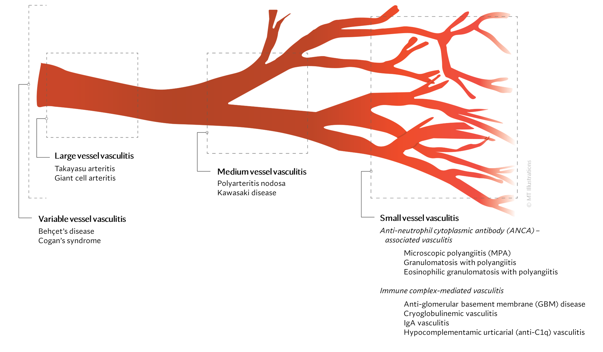
Vasculitis: What this website is all about!

Vasculitis Frequently Asked Questions

  • What causes vasculitis?
  • What is going to happen to me?
  • Is vasculitis curable?
  • Is vasculitis hereditary?
  • Does diet affect vasculitis?
  • Will my vasculitis return?
  • How should I guard against the occurrence of a disease flare?
  • Why do I have to have bloodwork checked frequently?

What causes vasculitis?

The causes of most forms of vasculitis remain unknown. Infections are strongly suspected of playing a role in in forms such as the association of hepatitis B (a virus) and polyarteritis nodosa, and hepatitis C (another virus) and cryoglobulinemic vasculitis. Bacterial infections have been suspected of playing a possible role in granulomatosis with polyangiitis (GPA, formerly known as Wegener’s) which is the reason that some patients with GPA that is limited to the upper respiratory tract are treated only with an antibiotic, Bactrim (trimethoprim/sulfamethoxazole). A general theory that applies to many types of vasculitis is that the disease results from the occurrence of a particular infection in a person whose genes (and other factors) make him/her susceptible to developing vasculitis.

What is going to happen to me?

The course of vasculitis is often difficult to predict. Some types of vasculitis may occur only once and do not return. Other types are prone to recurrences. For all patients with vasculitis, it is essential to be evaluated by physicians who are experienced in the treatment of these diseases. Vasculitis is treatable, and many patients achieve remissions through treatment. It is important to balance the types of medications necessary to control the disease and the risk of side effects that those medicines often bring. A primary aim of several ongoing new studies in vasculitis is to find drugs that help maintain remission.

Is vasculitis curable?

Most forms of vasculitis are treatable if detected early enough, before substantial organ damage has occurred. While often effective, however, the treatments remain imperfect and require improvement. Further research is needed in all forms of vasculitis. Greater knowledge of these diseases will lead to better treatments and, some day, to cures.

Will my children or other family members get it?

Vasculitis is not contagious. One cannot acquire vasculitis from contact with a vasculitis patient. In addition, despite the fact that genes probably play a role in susceptibility to some forms of vasculitis, it is unusual for vasculitis to occur in more than one member of the same family. Thus, vasculitis is not a heritable disorder. All of these points illustrate the fact that the causes of vasculitis are complex. In all likelihood, patients develop vasculitis because of the simultaneous occurrence of multiple risk factors, most of which remain poorly understood.

Does diet affect vasculitis?

This is one of the most commonly-asked questions by patients with vasculitis. All patients want to do whatever is within their power to help treat their disease. Unfortunately, there is presently no evidence that a person’s diet affects susceptibility to vasculitis, or that consuming or avoiding certain foods or beverages affects the course of the disease. In general, we advocate eating a balanced healthy diet rich in protein and vegetables. Avoidance of excessive empty calories, processed foods, and sugars may be very important, particularly in patients on steroids who are at risk for weight gain.

Will my vasculitis return?

After patients achieve remission from their vasculitis, it is logical for them to wonder if their disease will ever return. The answer, which is often difficult to give with certainty, depends in large part on the patient’s specific type of vasculitis. For example, some types of vasculitis, such as Henoch-Schönlein purpura (HSP) or vasculitis caused by a medication, are often self-limited and resolve on their own. Other forms of vasculitis (e.g., Buerger’s disease, a disease strongly associated with cigarette smoking) resolve with institution of the definitive treatment: smoking cessation.

However,  other forms of vasculitis behave less predictably and never come back in some patients but recur frequently in others. Granulomatosis with polyangiitis (GPA), giant cell arteritis (GCA), Takayasu arteritis, microscopic polyangiitis, and many other types of vasculitis fall into the category of diseases that have periods of quiescence and periods of flare. Disease flares in vasculitis can be mild (rash, minor joint pains) or severe (renal failure, skin ulcers). Flares may occur if medications are discontinued or dosage is lowered. Flare may occur in the context of infection. Often the reason for disease flare is unknown.

At the present time, the ability of doctors to predict who will suffer disease flares and who will maintain in long-term remissions (or be cured) needs refinement. Progress in this area will come through research.

How should I guard against the occurrence of a disease flare?

We believe that several points are worth keeping in mind:

First, the symptoms of flares are usually very similar those experienced at the onset of disease. If headaches signaled the beginning of giant cell arteritis, then the recurrence of headaches may indicate a disease flare. If leg ulcers began as painful red lumps on the leg the first time, then the return of painful red lumps may mean that vasculitis is back. Patients must become experts about their own manifestations of vasculitis so that they can recognize them immediately, consult their doctors, and begin appropriate treatment before serious damage occurs.

Second, we believe that patients truly know and understand their own bodies. It is important to discuss new or changing symptoms with your physicians. Together, patients and physicians can determine if new symptoms truly represent a vasculitis flare or if the cause is something equally as likely (medication side effect, infection, or other common medical issues).

Finally, because vasculitis treatments require careful monitoring by doctors, patients should discuss any changes in treatment with their physicians. Increasing or decreasing medications without consulting a physician may lead to trouble.

Why do I have to have bloodwork checked frequently?

Blood tests are helpful to monitor for the return of vasculitis by keeping a watchful eye on important parameters such as kidney function, liver tests, and markers of inflammation (ESR and CRP). Blood tests are also very important to ensure that medications are not causing any side effects such as liver irritation or low blood counts.

How often should my blood be checked?

This depends on the specific medicine or medicines that you take. Patients on cyclophosphamide (Cytoxan) should have their counts checked every week. Patients on most other kinds of medications used to treat vasculitis (Methotrexate, Azathioprine) usually only need to have their blood work checked monthly. If some laboratory tests are abnormal or nearly so, then more frequent monitoring may be required.

What type of tests do we check?

Regardless of the type of vasculitis and the exact type of medication that a patient takes, similar types of tests are monitored. These tests are:

  1. a complete blood count;
  2. tests of kidney function including a urinalysis; and
  3. liver function tests.

The table below outlines the importance behind checking each of these tests.

Type of TestWhat should be checkedWhy?
Complete Blood Count (“CBC”)
  • White blood cells (WBC)
  • Platelets
  • Hematocrit
  • Low WBC count may lead to infections.
  • Low platelets may cause bleeding.
  • Low hematocrit means insufficient oxygen-carrying capacity of the blood.
Kidney Function
  • Creatinine
  • Blood Urea Nitrogen (BUN)
  • High creatinine and BUN indicate that the kidneys are not performing their blood-cleansing function properly.
Urinalysis
  • Protein Level
  • Red Blood Cells
  • Normal urinalyses have no protein and no blood.
  • The presence of protein and/or blood in the urine may indicate active vasculitis in the kidneys (or damage to the bladder from cyclophosphamide).
Liver Function
  • Albumin
  • Aspartate aminotransferase(AST)
  • Alanine aminotransferase (ALT)
  • Often a good indication of overall health.
  • Elevated AST/ALT levels indicate inflammation in the liver (usually caused by medications).

Rheumatoid Vasculitis

  • First Description
  • Who gets Rheumatoid Vasculitis (the “typical” patients)?
  • Classic symptoms of Rheumatoid Vasculitis
  • What causes Rheumatoid Vasculitis?
  • How is Rheumatoid Vasculitis diagnosed?
  • Treatment and Course of Rheumatoid Vasculitis
  • What’s new in Rheumatoid Vasculitis?

First Description

Rheumatoid Vasculitis (RV) is an unusual complication of longstanding, severe rheumatoid arthritis. The active vasculitis associated with rheumatoid disease occurs in about 1% of this patient population.

RV is a manifestation of “extra-articular” (beyond the joint)rheumatoid arthritis and involves the small and medium-sized arteries in the body. In many of its disease features, RV resembles polyarteritis nodosa.

Other common extra-articular manifestations of rheumatoid arthritis, such as inflammation in the sac surrounding the heart (pericarditis), inflammation in the lining of the lungs (pleuritis), and interstitial lung disease (resulting in fibrosis or scarring of the lungs).

Who gets Rheumatoid Vasculitis? A typical patient

RV can affect a person from any ethnic background, either gender, and from any age group. However, more often than not, the typical patient has long-standing rheumatoid arthritis with severe joint deformities from the underlying arthritis. Although the arthritis has usually led to significant joint damage, at the onset of RV the joint disease is paradoxically quiet.

Figure: Patient with joint damage from rheumatoid arthritis. Note the bulbous swelling of some knuckles and lateral (ulnar) deviation of the fingers.

Classic symptoms of Rheumatoid Vasculitis

RV has many potential signs and symptoms. The manifestations of RV can involve many of the body’s different organ systems, including but not limited to the skin, peripheral nervous system (nerves to the hands and feet) , arteries of the fingers and toes causing digital ischemia, and eyes with scleritis. Scleritis (inflammation of the white part of the eye) commonly occurs in the setting of RV. This ocular complication requires urgent treatment with immunosuppressive medications.

Figure: Digital ischemia – this image shows a blood flow deficiency in the tip of the finger caused by an obstruction of the digital artery.

Figure: Scleritis – Inflammation of the sclera (the white of the eye) causing redness, light sensitivity and pain.

In addition, generalized symptoms such as fever and weight loss are common.

As is true with other forms of vasculitis that involve the skin, cutaneous lesions can erupt on various areas of the body in RV, with a predilection for the lower extremities. Typical findings include ulcers concentrated near the ankles.

Figure: Cutaneous ulcer – an open skin sore caused by an obstruction of the small blood vessels in the superficial ulcers or obstruction of medium vessels in a deeper ulcer.

Small nail fold infarcts (small spots around fingernail) can

occur in rheumatoid arthritis

but these do not necessarily signify the presence of systemic vasculitis and do not necessitate a change in rheumatoid arthritis treatment.

Nerve damage can cause foot or wrist drop, known in medical terminology as “mononeuritis multiplex”. The images below show a patient with a right wrist drop and a patient with right foot drop. This condition, which may be significantly disabling, is often preceded by a change in sensation in the same area (numbness, tingling, burning, or pain). These abnormal sensations can progress to muscle weakness, focal paralysis, and eventually to muscle wasting. Recovery from this condition, caused by nerve infarction, can take months. In some cases, recoveries from mononeuritis multiplex are incomplete.

Figures of drop wrist and drop foot (courtesy of the University of North Carolina)

(Video of drop foot viewable on our Microscopic Polyangiitis page under classic symptoms.)

Laboratory Tests

Most laboratory findings in RV – for example, elevations in the erythrocyte sedimentation rate or C-reactive protein – are non-specific, and reflect the presence of a generalized inflammatory state. Hypocomplementemia, anti-nuclear antibodies (ANA), and atypical anti-neutrophil cytoplasmic antibodies (ANCA) are common. Rheumatoid factor levels are usually extremely elevated. However, there is no definitive laboratory test for RV short of a tissue biopsy. The diagnosis must usually be made using a combination of history, physical examination, pertinent laboratory investigations, specialized testing (e.g., nerve conduction studies), and sometimes a tissue biopsy.

Because the treatment implications for RV are major, any diagnostic uncertainty must be met with definitive approaches to establishing the diagnosis. This usually involves biopsy of an involved organ. Deep skin biopsies (full-thickness biopsies that include some subcutaneous fat) taken from the edge of ulcers are very useful in detecting medium-vessel vasculitis. Nerve conduction studies help identify involved nerves for biopsy. Muscle biopsies (e.g., of the gastrocnemius muscle) should be performed at the same time as nerve biopsies, to increase the chance of finding changes characteristic of vasculitis. Imaging studies have no consistent role in the evaluation of RV, although sometimes angiography of the gastrointestinal tract is useful.

What Causes Rheumatoid Vasculitis?

The cause of RV is unknown, but given the prominence of immune components and the pathologic changes in involved blood vessels, an auto-immune process is suggested.

How is Rheumatoid Vasculitis diagnosed?

Most laboratory findings in RV – for example, elevations in the erythrocyte sedimentation rate or C-reactive protein are non-specific, and reflect the presence of a generalized inflammatory state. Hypocomplementemia, anti-nuclear antibodies (ANAs), and atypical anti-neutrophil cytoplasmic antibodies (atypical ANCAs) are common. Rheumatoid factor levels are extremely elevated, as a rule. However, there is no definitive laboratory test for RV short of a tissue biopsy. The diagnosis must usually be made by the combination of history, physical examination, pertinent lab work, other specialized testing (e.g., nerve conduction studies), and sometimes even a tissue biopsy is required.

The diagnosis of RV should be considered in any rheumatoid arthritis patient who develops new constitutional symptoms, skin ulcerations, decreased blood flow to the fingers or toes, symptoms of a sensory or motor nerve dysfunction (numbness, tingling, focal weakness); or any inflammation of the lining around the heart or lungs (pericarditis or pleurisy/pleuritis).

Patients with a history of joint-destructive rheumatoid arthritis are at an increased risk for infection. Therefore, when a rheumatoid arthritis patient presents with a new onset of non-specific systemic complaints an infection must first be eliminated. Patients with rheumatoid arthritis typically have immune systems that are disordered from previous immunosuppression and underlying disease (e.g., joint damage). This patient population, therefore, is at higher risk of infection.

The differential diagnosis of RV includes:

  • Cholesterol embolization syndromes, in which a piece of cholesterol breaks off of a plaque, may cause digital ischemia (blood flow obstruction to a finger or toe), and a host of other symptoms that mimic vasculitis.
  • Diabetes mellitus is another major cause of mononeuritis multiplex, but multiple mononeuropathies occurring over a short period of time are unusual in diabetes.
  • Many clinical features of RV mimic those of polyarteritis nodosa, cryoglobulinemia, and other forms of necrotizing vasculitis. Therefore they too should be considered in this setting.

Because the treatment implications for RV are major, any diagnostic uncertainty must be met with a definitive approach to establishing the diagnosis. As alluded to earlier, this usually involves the biopsy of an involved organ. Deep skin biopsies (full-thickness biopsies that include some subcutaneous fat) taken from the edge of ulcers are very useful in detecting medium-vessel vasculitis. Nerve conduction studies help identify involved nerves for biopsy. Muscle biopsies (e.g., of the gastrocnemius muscle) should be performed at the same time as nerve biopsies, to increase the chance of finding changes characteristic of vasculitis. Imaging studies have no consistent role in the evaluation of RV, although sometimes angiography of the gastrointestinal tract is useful.

Normally, the cells of the blood vessel wall would be fewer in number (less thick) and the lumen (larger red area) would be larger. The arrow points (Figure 6, left) to an inflamed blood vessel found on a muscle biopsy. The globular pink areas are muscle fibers.

Treatment and Course of Rheumatoid Vasculitis

Therapy should reflect the severity of organ involvement. Prednisone or other steroid therapies are often the first line of treatment. Optimizing treatment of the underlying rheumatoid arthritis is also essential, therefore medications such as methotrexate or tumor necrosis factor inhibitors may be employed. In the setting of impending damage to major organs such as the eyes, a peripheral nerve, the gastrointestinal tract, or of a severe skin ulceration, cyclophosphamide is usually warranted.

What’s New in Rheumatoid Vasculitis?

Compared to other forms of vasculitis, there has been relatively little research in recent years on the specific entity of RV. The lack of similarity in available reports on RV and discrepancies in case definitions have created challenge to building standard approaches to the diagnosis and treatment of this condition. There is some evidence that the incidence of RV has decreased over the past several decades, perhaps because of better treatment of the underlying rheumatoid arthritis.

Meet Our Team

Vasculitis Center Doctors

Duvuru Geetha, MD

Professor of Clinical Medicine

Dr. Geetha is a Professor of Medicine in the Division of Nephrology. A graduate of Madras Medical College, India, she completed Internal Medicine training in U.K. She did her Internal Medicine Residency at York, PA and Nephrology fellowship at Johns Hopkins Bayview Medical Center. She has been on Hopkins faculty since 1998. She is a member of Royal College of Physicians (U.K.), American Society of Nephrology, American Society of Transplantation and a consultant for the vasculitis foundation. She is a member of the Miller Coulson Academy of Clinical Excellence at Hopkins. Her clinical interests include renal disease in vasculitis patients with a focus on ANCA associated vasculitis and Henoch-Schonlein Purpura. She does clinical and translational research in vasculitis with a focus on ANCA associated vasculitis and renal disease.

Brendan Antiochos, MD

Assistant Professor of Medicine

Dr. Antiochos is a graduate of Dartmouth College and Dartmouth Medical School. He completed internal medicine residency at Oregon Health & Science University, then rheumatology fellowship at Johns Hopkins, before joining the faculty here in 2014. Dr. Antiochos assumed the role of Director for the vasculitis center in 2022. In addition to seeing patients in the vasculitis center, Dr. Antiochos performs laboratory-based research on autoimmune diseases. His laboratory work focuses on activation of the innate immune system and the discovery of novel autoantibodies.

Philip Seo, MD

Associate Professor of Medicine

A graduate of Harvard College and the College of Physicians and Surgeons at Columbia University, Dr. Seo completed his Internal Medicine training as a member of the Osler Medical Service at the Johns Hopkins Hospital. Since then, he has worked at Johns Hopkins in several capacities, including as a hospitalist at Johns Hopkins Bayview Medical Center, and as an Assistant Chief of Service of the Department of Medicine at the Johns Hopkins Hospital, before joining the Division of Rheumatology. His research interests are the assessment and treatment of ANCA-associated vasculitides, including Churg Strauss Syndrome, Wegener’s Granulomatosis, and Microscopic Polyangiitis.

David B. Hellmann, MD

Aliki Perroti Professor of Medicine

Dr. Hellmann is the Chairman of the Department of Medicine and Vice Dean at The Johns Hopkins Bayview Medical Center, and the Aliki Perroti Professor of Medicine. A graduate of Yale University and Johns Hopkins Medical School, Dr. Hellmann received his Internal Medicine training on the Osler Service at Hopkins, and trained in Rheumatology at the University of California, San Francisco. He has been on the Johns Hopkins faculty since 1986.

Desh Nepal, MD

Assistant Professor of Medicine

Michael Cammarata, MD

Assistant Professor of Medicine

Dr. Cammarata is a graduate of The College of William & Mary. He attended Eastern Virginia Medical School and completed his residency in Internal Medicine at the University of California San Francisco. He returned to the east coast for rheumatology fellowship at Johns Hopkins, joining faculty in 2024. He is RhMSUS certified in musculoskeletal ultrasound, and also practices general medicine as a hospitalist at Johns Hopkins Hospital. 

Collaborators

Vasculitis can involve virtually any organ system within the body. Hence, our Vasculitis Center maintains close collaborative relationships with experts from other specialties. The Vasculitis Center includes collaborators from several medical disciplines who help provide the highest level of care for our patients. They have extensive experience managing vasculitis within their subspecialty and work closely with the Physicians in the Vasculitis Center to provide comprehensive care for our patients:

Otolaryngology (ENT):

Our ENT team includes specialists in inflammatory sinus disease, sensorineural hearing loss, and chronic middle ear disease. We are pleased to also have a Doctor of Audiology, Dr. Dinkes, who specializes in inflammatory process on our team as well.

  • Dr. Jean Kim (sinus disease, middle ear manifestations)
  • Dr. Alexander Hiller (upper airway disease)
  • Dr. Roni Dinkes (audiology)

Neuro-ophthalmology:

  • Dr. Andrew Carey

Endocrinology / Osteoporosis:

  • Dr. Han Na Kim

Resources

Living with vasculitis can be challenging at times due to the complex nature of the disease and therapies. Also, vasculitis in the general population is quite rare, especially compared to other common medical conditions, such as diabetes and hypertension (high blood pressure). This can make patients with vasculitis feel misunderstood or alone. It can be very helpful for individuals with vasculitis to connect with other patients and their families.

If you have vasculitis, you are not alone. There is a strong community of patients and physicians to support you.

  • Vasculitis Foundation (USA) – www.vasculitisfoundation.org
  • Vasculitis UK – www.vasculitis.org.uk/
  • Vasculitis Foundation Canada – www.vasculitis.ca/
  • American Behcet’s Disease Association
  • Churg-Strauss Syndrome Association
  • Cryoglobulinemia Home Page
  • Arthritis Foundation
  • The American College of Rheumatology
  • European Vasculitis Study Group: EUVAS Homepage

Types of Vasculitis

There are approximately 20 different disorders that are classified as “vasculitis”. “Angiitis” and “Arteritis” are both synonyms for vasculitis, literally meaning “inflammation within blood vessels” or “inflammation in arteries.”  Because there are so many types of vasculitis, the group is sometimes referred to in the plural: vasculitides (pronounced “vas que lit’ i deez”).

There are many different types of diseases that belong to this category. Although the diseases are similar in some ways, they often differ with respect to which organs are affected, which medications are used to treat them, and other characteristics.

In general, vasculitis can be grouped based on the size of the blood vessels that are
mainly affected. However, it is important to keep in mind that any sized blood vessels can be
involved.

Large vessel vasculitis (LVV)
LVV is characterized by inflammation of the largest-sized blood vessels of the body such
as the aorta, major arteries that deliver blood to distant parts of the body. They also include
large veins that deliver blood back to the heart. These are:

  • Takayasu arteritis
  • Giant cell arteritis

Medium vessel vasculitis (MVV)
MVV is inflammation of the medium-sized blood vessels including arterioles and smaller
veins.

  • Polyarteritis nodosa
  • Kawasaki disease

Small vessel vasculitis (SVV)
SVV affects the smallest blood vessels of the body called capillaries and venules. SVV
involves inflammation mediated by autoantibodies (antibodies are molecules that usually coats
bacteria and viruses; however, in autoimmune disease, they bind to cells and proteins normally
found in the body, hence the name, autoantibodies). These antibodies can deposit in small vessels
causing restriction of blood flow to tissue.
They can be of two types:

(1) Anti-neutrophil cytoplasmic antibody (ANCA)-associated vasculitis (AAV)

  • Microscopic polyangiitis (MPA)
  • Granulomatosis with polyangiitis (GPA)
  • Eosinophilic granulomatosis with polyangiitis (EGPA)


(2) Immune complex small vessel vasculitis

  • Anti-glomerular basement membrane
  • Cryoglobulinemic vasculitis
  • IgA vasculitis
  • Hypocomplementamic urticarial vasculitis (anti-C1q vasculitis)

Variable vessel vasculitis (VVV)
This type of vasculitis can involve inflammation of blood vessels of any size.

  • BehÇet’s disease
  • Cogan’s syndrome

Others:

Single-organ vasculitis (SOV):
SOV is inflammation affecting the blood vessels that feed a single organ, such as the brain or skin.

Vasculitis associated with another autoimmune disease:
Vasculitis can occur in conjunction with other autoimmune diseases, such as lupus or sarcoidosis.

Vasculitis associated with other diseases:
Other diseases, such as infections and cancer, can cause systemic vasculitis as well.
Vasculitis as a side effect of drugs:
Certain drugs, such as hydralazine and cocaine, can cause vasculitis.

  • « Go to Previous Page
  • Go to page 1
  • Go to page 2
  • Go to page 3
  • Go to page 4
  • Go to Next Page »

Primary Sidebar


donate to the Johns Hopkins Vasculitis Center

All information contained within the Johns Hopkins Vasculitis Center website is intended for educational purposes only. Visitors are encouraged to consult other sources and confirm the information contained within this site. Consumers should never disregard medical advice or delay in seeking it because of something they may have read on this website.

Footer

Johns Hopkins Rheumatology

  • Johns Hopkins Rheumatology
  • Johns Hopkins Arthritis Center
  • Johns Hopkins Lupus Center
  • Johns Hopkins Lyme Disease Research Center
  • Johns Hopkins Myositis Center
  • Johns Hopkins Scleroderma Center
  • Johns Hopkins Sjögren’s Syndrome Center

Connect With Us

  • Facebook
  • Twitter
  • YouTube
U.S. News and World Report Rankings Badge

Johns Hopkins Medicine Johns Hopkins Medicine logo
Language Assistance Available:
  • Español
  • American Sign Language
  • עִברִי
  • אידיש
  • አማርኛ
  • 繁體中文
  • Français
  • Tagalog
  • Русский
  • Português
  • Italiano
  • Tiếng Việt
  • Ɓàsɔ́ɔ̀-wùɖù-po-nyɔ̀
  • Igbo asusu
  • èdè Yorùbá
  • বাংলা
  • 日本語
  • 한국어
  • Kreyòl Ayisyen
  • العربية
  • Deutsch
  • Polski
  • Ελληνικά
  • ગુજરાતી
  • ภาษาไทย
  • اُردُو
  • فارسی
  • हिंदी
  • Deitsch
  • ខ្មែរ
  • မြန်မာ
Contact & Privacy Information
Price Transparency
Notice of Privacy Practices Privacy Statement
Terms & Conditions of Use Non-Discrimination Notice
Manage Cookie Preferences
Copyright © 2025 Johns Hopkins Rheumatology