• Skip to primary navigation
  • Skip to main content
  • Skip to primary sidebar
  • Skip to footer

Johns Hopkins Vasculitis Center

Show Search
Hide Search
  • About Our Center
    • Appointments and Directions
    • Meet Our Team
    • Support Our Center
  • What is Vasculitis?
    • Types of Vasculitis
    • Causes of Vasculitis
    • Symptoms of Vasculitis
    • Diagnosing Vasculitis
  • Vasculitis Treatments
    • Prednisone
    • Avacopan (Tavneos®)
    • Apremilast (Otezla®)
    • Azathioprine
    • Colchicine
    • Cyclophosphamide (Cytoxan)
    • Dapsone
    • Supplemental Immunoglobulin (IVIG/SCIG)
    • Leflunomide
    • Mepolizumab (Nucala®)
    • Methotrexate (MTX)
    • Mycophenolate
    • Rituximab
    • Sarilumab (Kevzara®)
    • TNF Inhibitors
    • Tocilizumab (Actemra®)
  • Vasculitis Research
  • Resources
Home / Search for "search.html/whatis/cryoglobulinemia.html/treatments/methotrexate.html"

Search Results for: search.html/whatis/cryoglobulinemia.html/treatments/methotrexate.html

Takayasu’s Arteritis

  • First Description
  • Who gets Takayasu’s Arteritis (the “typical” patients)?
  • Classic symptoms of Takayasu’s Arteritis
  • What causes Takayasu’s Arteritis?
  • How is Takayasu’s Arteritis diagnosed?
  • Treatment and Course of Takayasu’s Arteritis
  • What’s new in Takayasu’s Arteritis?
  • In medical terms, by David Hellmann, M.D.

First Description

The first case of Takayasu’s arteritis was described in 1908 by Dr. Mikito Takayasu at the Annual Meeting of the Japan Ophthalmology Society. Dr. Takayasu described a peculiar “wreathlike” appearance of blood vessels in the back of the eye (retina). Two Japanese colleagues at the same meeting reported similar eye findings in patients whose wrist pulses were absent. It is now known that the blood vessel malformations that occur in the retina are a response (new blood vessel growth) to arterial narrowings in the neck, and that the absence of pulses noted in some patients occur because of narrowings of blood vessels to the arms. The eye findings described by Dr. Takayasu are rarely seen in patients from North America.

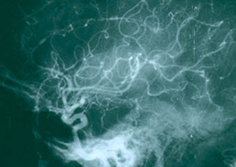
Pictured below is a close–up view of an angiogram of the left vertebral and subclavian arteries in a patient with Takayasu’s arteritis. Note the narrowing and irregularities that occur at several sites, and the “corkscrew” configuration of one vessel segment near the junction of the two arteries. These changes, caused by inflammation in the blood vessel wall, sometimes cause complete blockage of the artery.

Takayasu’s arteritis is occasionally called “pulseless disease”, because of the difficulty in detecting peripheral pulses that sometimes occurs as a result of the vascular narrowings.

Who gets Takayasu’s Arteritis?

The “typical” patient with Takayasu’s arteritis is a woman under the age of 40. There is a 9:1 female predominance in this disease. Although the disease has a worldwide distribution, it appears to occur more often in Asian women.

Takayasu’s arteritis is a rare disease. The best estimates of the disease frequency suggest that 2 or 3 cases occur each year per million people in a population.

Classic Symptoms of Takayasu’s Arteritis

Takayasu’s arteritis is a chronic inflammatory condition that affects the largest blood vessel in the body (the aorta) and its branches. Thus, the complications of Takayasu’s arise directly or indirectly from damage to these blood vessels. The vasculitides are classified according to the size of blood vessel involved. Takayasu’s is the classic “large vessel” vasculitis.

Pictured below is a normal aortic arch on the left, with narrow, smooth blood vessels. On the right is an example of an abnormal aortic arch in a patient with Takayasu’s, with obvious dilation of the ascending aorta on the left side of the picture.

Clinicians divide Takayasu’s arteritis into two phases: 1) a systemic phase; and 2) an occlusive phase. Although these two phases are not always distinct (i.e., patients may have features of both phases at the same time), this division is a useful way of thinking about the disease.

In the systemic phase, patients have symptoms and signs of an active inflammatory illness. These may include “constitutional symptoms” (fever, fatigue, weight loss), arthritis, and non-specific aches and pains. There may be tenderness overlying affected arteries. Most patients have elevations of the erythrocyte sedimentation rate during the systemic phase.

The systemic phase is succeeded by the occlusive phase, during which patients begin to develop symptoms caused by the narrowing of affected arteries. These may include pain in the limbs that occurs during repetitive activities (“claudication”), such as pain in the arm that occurs while using a handsaw or pain in the calves brought on by walking. The symptoms also include dizziness upon standing up, headaches, and visual problems. During the occlusive phase, affected blood vessels may be narrowed to such an extent that the normal arterial pulsations (“pulses”) in the neck, elbow, wrist, or lower extremities cannot be felt. Using a stethoscope, one may also hear “bruits” (pronounced ‘brew eez’), a harsh, “whooshing” sounds made by the flow of blood through abnormally narrowed vessels. High blood pressure is common, but blood pressures taken in the arms may be read as falsely low if there is a narrowing of an artery high up in the arm. With some patients, it is not possible to get accurate blood pressure readings in the arms. Using an ophthalmoscope, a physician may observe characteristic malformations of blood vessels that occur in advanced cases of Takayasu’s arteritis.

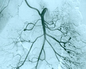
Although the lung involvement in Takayasu’s is frequently overshadowed by involvement of systemic large blood vessels, the pulmonary arteries may also be affected in this disorder. Pictured below is a pulmonary angiogram demonstrating beading and cut–off lesions of the right pulmonary arteries, and a large aneurysm of the left pulmonary artery.

What Causes Takayasu’s Arteritis?

The cause of Takayasu’s arteritis is not known. Some evidence suggests that an infection of some kind — viral, bacterial, or other — occurring in a person with other predisposing factors (such as the correct genes), may lead to this disease. This is an attractive hypothesis, but definitive evidence for it is lacking.

How is Takayasu’s Arteritis Diagnosed?

Making the diagnosis of Takayasu’s arteritis can be extremely difficult. Unfortunately it is very common for the disease to smoulder in the walls of large blood vessels for years, causing only non-specific symptoms associated with the systemic phase of the illness (or no symptoms), until a major complication results. These major complications may include dilation of the aorta with “stretching” of the aortic valve in the heart; critically reduced blood flow to an arm or leg; a stroke caused by high blood pressure in vessels of the brain, and many others.

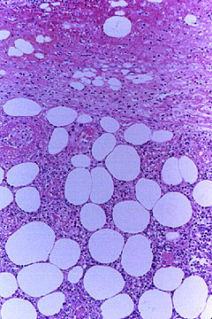
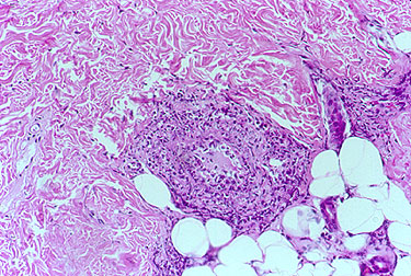
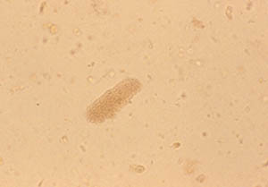
Once the diagnosis is suspected, it is usually confirmed by a radiographic procedure such as an angiogram or a magnetic resonance imaging study demonstrating significant large artery disease consistent with Takayasu’s. In some cases in which blood vessel damage is so severe as to necessitate surgery to repair the aortic valve, the aorta, or some other large blood vessel, physicians are able to make unequivocal diagnoses by looking at tissue from the involved blood vessels under the microscope. Takayasu’s arteritis is pathologically indistinguishable from giant cell arteritis. In both, destruction of the blood vessel wall and giant cells are frequently present.

Pictured below is an example of large artery involvement in Takayasu’s arteritis. Magnetic resonance imaging study of the aorta and large blood vessels of the upper extremities, showing a large aneurysm of the ascending aorta, blockage of the right axillary artery (note the interruption of blood flow near the shoulder on the left of the figure), and many narrowings of the left subclavian artery (on the right of the figure).

Treatment and Course of Takayasu’s Arteritis

The great majority of patients with Takayasu’s arteritis respond to prednisone. The usual starting dose is approximately 1 milligram per kilogram of body weight per day (for most people, this is approximately 60 milligrams a day). Because of the significant side–effects of long-term high–dose prednisone use, the starting dose is tapered over several weeks to a dose that the physician feels is tolerable for the patient.

For long–term treatment in addition to prednisone (as “steroid sparing agents”), methotrexate, azathioprine, and even cyclophosphamide are sometimes used. There have been few studies of the use of these medications in this disease.

What’s New in Takayasu’s Arteritis?

One of the biggest problems confronting Takayasu’s patients and the physicians who care for them is determining how active the disease is. This can be an exceptionally challenging problem. The erythrocyte sedimentation rate (ESR) probably remains the most reliable marker of disease activity, but even this test is not helpful in a sizeable number of patients who have active arterial inflammation but normal ESRs. Because the treatments for Takayasu’s arteritis may be associated with substantial side–effects, we need more accurate means of gauging disease activity.

To this end, a study conducted by the International Network for the Study of Systemic Vasculitides (“INSSYS”) may be helpful. Investigators from INSSYS, which includes more than 300 physicians, scientists, and other experts in vasculitis from more than 50 different medical centers across the world, have been conducting a “Surrogate Markers Study” for the past several years. In this study, the investigators examine blood specimens from patients with vasculitis for the purpose of identifying proteins and other molecules whose presence indicates ongoing inflammation. Improved understanding of these diseases at a molecular level may permit more rational use of treatments in the future.

In medical terms, by David Hellmann, M.D.

A discussion of Takayasu’s Arteritis written in medical terms by David Hellmannn, M.D. (F.A.C.P.), The Johns Hopkins Vasculitis Center, for the Rheumatology Section of the Medical Knowledge Self-Assessment Program published and copyrighted by the American College of Physicians (Edition 11, 1998). The American College of Physicians has given us permission to make this information available to patients contacting our Website.

Takayasu’s arteritis is a granulomatous vasculitis chiefly of young women that involves the aorta and its major branches. Patients can present initially with obscure systemic symptoms such as fever of unknown origin or more commonly with symptoms and signs of large vessel vasculitis such as hypertension from renal artery stenosis, aortic regurgitation from aortitis, or stroke from carotid artery occlusion. Bruits and diminished or absent pulses are the most reliable signs. Anemia and elevated ESR accompany active disease. Diagnosis is confirmed by angiography showing stenosis and dilation of the aorta, its branches, or both. Thickening of the aortic wall detectable by MRI or ultrasonography can precede angiographic changes. Prednisone is effective for the systemic symptoms and can thwart progression of the vasculitis. Methotrexate may be an effective corticosteroid-sparing agent. Angioplasty alleviates renal artery stenosis about half the time. When needed, vascular bypass procedures and aortic valve replacement usually work well if deferred until the disease is inactive. Estimating disease activity is difficult but is based on systemic symptoms, anemia, ESR, progression of lesions, and pathology (when available).

About Our Center

About Our Center

Our center is composed of dedicated physicians, research coordinators and patient care coordinators who serve patients with vasculitis. Many patients who receive these diagnoses have never previously heard the term “vasculitis” or met other patients with the same condition. The vasculitis syndromes (known together as “the vasculitides”) are a group of diseases that can affect every organ system, and occur in people of all ages, genders and backgrounds. Because these diseases are relatively rare and can present in many different ways, the diagnosis of vasculitis is often difficult to reach, and many patients suffer a period of uncertainty prior to finally arriving at a diagnosis of vasculitis. In our mission of serving all patients with vasculitis, we consider the provision of clear and accurate information to be one of our most important responsibilities.

At this Website you will find:

  1. explanations of vasculitis in lay terms
  2. specific discussions of individual diseases
  3. a review of the common therapies for vasculitis
  4. answers to commonly asked questions
  5. information on how to make an appointment to be seen in the Johns Hopkins Vasculitis Center
  6. information about ongoing research at the Johns Hopkins Vasculitis Center
  7. ways in which you can contribute to advancing research and progress in vasculitis.

Please note that this Website is sponsored through the generosity of various friends of the Johns Hopkins Vasculitis Center. We update the Vasculitis Center Website regularly and strive to provide solid, usable information on various types of vasculitis, treatments, and support resources.

Thank you for visiting our Website. We hope you will find it accessible and useful as you learn about these challenging diseases.

Yours truly,

Brendan Antiochos, MD
Assistant Professor of Medicine
Johns Hopkins University School of Medicine,
Division of Rheumatology
Director, The Johns Hopkins Vasculitis Center

Welcome to the Johns Hopkins Vasculitis Center

Welcome to the Johns Hopkins Vasculitis Center

Dear Vasculitis Center Website Visitor:

SEO

Welcome to the Johns Hopkins Vasculitis Center Website. This Website, maintained by the Physicians, Research Coordinators, and Patient Care Coordinators at our Center, is designed to provide information for patients with vasculitis in language that non-medical people can understand. We recognize that many patients with vasculitis have never heard of their disease before they became sick and that, owing to the relative rarity of some types of vasculitis, most physicians have little experience treating the disorders. Few support groups for vasculitis patients exist, and there is a shortage of literature about these diseases written for lay people. Consequently, most patients find reliable information about vasculitis difficult to come by.

At this Website you will find:

  1. explanations of vasculitis in lay terms
  2. specific discussions of individual diseases
  3. a review of the common therapies for vasculitis
  4. answers to commonly asked questions
  5. information on how to make an appointment to be seen in the Johns Hopkins Vasculitis Center
  6. information about ongoing research at the Johns Hopkins Vasculitis Center
  7. ways in which you can contribute to advancing research and progress in vasculitis.

Please note that this Website is sponsored through the generosity of various friends of the Johns Hopkins Vasculitis Center. We update the Vasculitis Center Website regularly and strive to provide solid, usable information on various types of vasculitis, treatments, and support resources.

Thank you for visiting our Website. We hope you will find it accessible and useful as you learn about these challenging diseases.

Yours truly,

Philip Seo, MD, MHS
Assistant Professor of Medicine
Johns Hopkins University School of Medicine,
Division of Rheumatology
Director, The Johns Hopkins Vasculitis Center

Rheumatoid Vasculitis

  • First Description
  • Who gets Rheumatoid Vasculitis (the “typical” patients)?
  • Classic symptoms of Rheumatoid Vasculitis
  • What causes Rheumatoid Vasculitis?
  • How is Rheumatoid Vasculitis diagnosed?
  • Treatment and Course of Rheumatoid Vasculitis
  • What’s new in Rheumatoid Vasculitis?

First Description

Rheumatoid Vasculitis (RV) is an unusual complication of longstanding, severe rheumatoid arthritis. The active vasculitis associated with rheumatoid disease occurs in about 1% of this patient population.

RV is a manifestation of “extra-articular” (beyond the joint)rheumatoid arthritis and involves the small and medium-sized arteries in the body. In many of its disease features, RV resembles polyarteritis nodosa.

Other common extra-articular manifestations of rheumatoid arthritis, such as inflammation in the sac surrounding the heart (pericarditis), inflammation in the lining of the lungs (pleuritis), and interstitial lung disease (resulting in fibrosis or scarring of the lungs).

Who gets Rheumatoid Vasculitis? A typical patient

RV can affect a person from any ethnic background, either gender, and from any age group. However, more often than not, the typical patient has long-standing rheumatoid arthritis with severe joint deformities from the underlying arthritis. Although the arthritis has usually led to significant joint damage, at the onset of RV the joint disease is paradoxically quiet.

Figure: Patient with joint damage from rheumatoid arthritis. Note the bulbous swelling of some knuckles and lateral (ulnar) deviation of the fingers.

Classic symptoms of Rheumatoid Vasculitis

RV has many potential signs and symptoms. The manifestations of RV can involve many of the body’s different organ systems, including but not limited to the skin, peripheral nervous system (nerves to the hands and feet) , arteries of the fingers and toes causing digital ischemia, and eyes with scleritis. Scleritis (inflammation of the white part of the eye) commonly occurs in the setting of RV. This ocular complication requires urgent treatment with immunosuppressive medications.

Figure: Digital ischemia – this image shows a blood flow deficiency in the tip of the finger caused by an obstruction of the digital artery.

Figure: Scleritis – Inflammation of the sclera (the white of the eye) causing redness, light sensitivity and pain.

In addition, generalized symptoms such as fever and weight loss are common.

As is true with other forms of vasculitis that involve the skin, cutaneous lesions can erupt on various areas of the body in RV, with a predilection for the lower extremities. Typical findings include ulcers concentrated near the ankles.

Figure: Cutaneous ulcer – an open skin sore caused by an obstruction of the small blood vessels in the superficial ulcers or obstruction of medium vessels in a deeper ulcer.

Small nail fold infarcts (small spots around fingernail) can

occur in rheumatoid arthritis

but these do not necessarily signify the presence of systemic vasculitis and do not necessitate a change in rheumatoid arthritis treatment.

Nerve damage can cause foot or wrist drop, known in medical terminology as “mononeuritis multiplex”. The images below show a patient with a right wrist drop and a patient with right foot drop. This condition, which may be significantly disabling, is often preceded by a change in sensation in the same area (numbness, tingling, burning, or pain). These abnormal sensations can progress to muscle weakness, focal paralysis, and eventually to muscle wasting. Recovery from this condition, caused by nerve infarction, can take months. In some cases, recoveries from mononeuritis multiplex are incomplete.

Figures of drop wrist and drop foot (courtesy of the University of North Carolina)

(Video of drop foot viewable on our Microscopic Polyangiitis page under classic symptoms.)

Laboratory Tests

Most laboratory findings in RV – for example, elevations in the erythrocyte sedimentation rate or C-reactive protein – are non-specific, and reflect the presence of a generalized inflammatory state. Hypocomplementemia, anti-nuclear antibodies (ANA), and atypical anti-neutrophil cytoplasmic antibodies (ANCA) are common. Rheumatoid factor levels are usually extremely elevated. However, there is no definitive laboratory test for RV short of a tissue biopsy. The diagnosis must usually be made using a combination of history, physical examination, pertinent laboratory investigations, specialized testing (e.g., nerve conduction studies), and sometimes a tissue biopsy.

Because the treatment implications for RV are major, any diagnostic uncertainty must be met with definitive approaches to establishing the diagnosis. This usually involves biopsy of an involved organ. Deep skin biopsies (full-thickness biopsies that include some subcutaneous fat) taken from the edge of ulcers are very useful in detecting medium-vessel vasculitis. Nerve conduction studies help identify involved nerves for biopsy. Muscle biopsies (e.g., of the gastrocnemius muscle) should be performed at the same time as nerve biopsies, to increase the chance of finding changes characteristic of vasculitis. Imaging studies have no consistent role in the evaluation of RV, although sometimes angiography of the gastrointestinal tract is useful.

What Causes Rheumatoid Vasculitis?

The cause of RV is unknown, but given the prominence of immune components and the pathologic changes in involved blood vessels, an auto-immune process is suggested.

How is Rheumatoid Vasculitis diagnosed?

Most laboratory findings in RV – for example, elevations in the erythrocyte sedimentation rate or C-reactive protein are non-specific, and reflect the presence of a generalized inflammatory state. Hypocomplementemia, anti-nuclear antibodies (ANAs), and atypical anti-neutrophil cytoplasmic antibodies (atypical ANCAs) are common. Rheumatoid factor levels are extremely elevated, as a rule. However, there is no definitive laboratory test for RV short of a tissue biopsy. The diagnosis must usually be made by the combination of history, physical examination, pertinent lab work, other specialized testing (e.g., nerve conduction studies), and sometimes even a tissue biopsy is required.

The diagnosis of RV should be considered in any rheumatoid arthritis patient who develops new constitutional symptoms, skin ulcerations, decreased blood flow to the fingers or toes, symptoms of a sensory or motor nerve dysfunction (numbness, tingling, focal weakness); or any inflammation of the lining around the heart or lungs (pericarditis or pleurisy/pleuritis).

Patients with a history of joint-destructive rheumatoid arthritis are at an increased risk for infection. Therefore, when a rheumatoid arthritis patient presents with a new onset of non-specific systemic complaints an infection must first be eliminated. Patients with rheumatoid arthritis typically have immune systems that are disordered from previous immunosuppression and underlying disease (e.g., joint damage). This patient population, therefore, is at higher risk of infection.

The differential diagnosis of RV includes:

  • Cholesterol embolization syndromes, in which a piece of cholesterol breaks off of a plaque, may cause digital ischemia (blood flow obstruction to a finger or toe), and a host of other symptoms that mimic vasculitis.
  • Diabetes mellitus is another major cause of mononeuritis multiplex, but multiple mononeuropathies occurring over a short period of time are unusual in diabetes.
  • Many clinical features of RV mimic those of polyarteritis nodosa, cryoglobulinemia, and other forms of necrotizing vasculitis. Therefore they too should be considered in this setting.

Because the treatment implications for RV are major, any diagnostic uncertainty must be met with a definitive approach to establishing the diagnosis. As alluded to earlier, this usually involves the biopsy of an involved organ. Deep skin biopsies (full-thickness biopsies that include some subcutaneous fat) taken from the edge of ulcers are very useful in detecting medium-vessel vasculitis. Nerve conduction studies help identify involved nerves for biopsy. Muscle biopsies (e.g., of the gastrocnemius muscle) should be performed at the same time as nerve biopsies, to increase the chance of finding changes characteristic of vasculitis. Imaging studies have no consistent role in the evaluation of RV, although sometimes angiography of the gastrointestinal tract is useful.

Normally, the cells of the blood vessel wall would be fewer in number (less thick) and the lumen (larger red area) would be larger. The arrow points (Figure 6, left) to an inflamed blood vessel found on a muscle biopsy. The globular pink areas are muscle fibers.

Treatment and Course of Rheumatoid Vasculitis

Therapy should reflect the severity of organ involvement. Prednisone or other steroid therapies are often the first line of treatment. Optimizing treatment of the underlying rheumatoid arthritis is also essential, therefore medications such as methotrexate or tumor necrosis factor inhibitors may be employed. In the setting of impending damage to major organs such as the eyes, a peripheral nerve, the gastrointestinal tract, or of a severe skin ulceration, cyclophosphamide is usually warranted.

What’s New in Rheumatoid Vasculitis?

Compared to other forms of vasculitis, there has been relatively little research in recent years on the specific entity of RV. The lack of similarity in available reports on RV and discrepancies in case definitions have created challenge to building standard approaches to the diagnosis and treatment of this condition. There is some evidence that the incidence of RV has decreased over the past several decades, perhaps because of better treatment of the underlying rheumatoid arthritis.

Granulomatosis with Polyangiitis

  • Who gets Granulomatosis with Polyangiitis (the “typical” patients)?
  • Classic symptoms of Granulomatosis with Polyangiitis
  • What causes Granulomatosis with Polyangiitis?
  • How is Granulomatosis with Polyangiitis diagnosed?
  • Treatment and Course of Granulomatosis with Polyangiitis
  • What’s new in Granulomatosis with Polyangiitis?
  • In medical terms, by David Hellmann, M.D.

Who gets Granulomatosis with Polyangiitis?

Granulomatosis with Polyangiitis is nearly equally distributed between the sexes, with a slight male predominance. Granulomatosis with Polyangiitis typically occurs in middle age, but is found in people of all ages. Although it is unusual for Granulomatosis with Polyangiitis to occur in childhood, it is not unusual for a Granulomatosis with Polyangiitis patient to be in his/her 70s or even 80s at the time of diagnosis.

Pictured below is a chest x–ray showing bilateral lung nodules in a 27 year old Indian man with Granulomatosis with Polyangiitis.

Pictured below is a CT scan from the same patient. The view is a cross–section through the patient’s lungs. The CT scan not only permits a better appreciation of the lesions’ size, it also detects more lesions.

Granulomatosis with Polyangiitis can affect virtually any site in the body, but it has a predisposition for certain organs. The classic organs involved in Granulomatosis with Polyangiitis are the upper respiratory tract (sinuses, nose, ears, and trachea [the “windpipe”]), the lungs, and the kidneys. Listed below are the organs commonly involved in Granulomatosis with Polyangiitis and the specific disease manifestation(s) in each organ.

  • Ear
  • Eye
  • Nose
  • Sinuses
  • Trachea
  • Lungs
  • Kidney
  • Skin
  • Joints
  • Nerves
  • Miscellaneous

Ear

Ear infections that are slow to resolve. Recurrent otitis media. Decrease in hearing.

Eye

Inflammation can occur in different parts of the eye. Inflammation in the white part of the eye is known as the sclera (“scleritis”). “Uveitis” is inflammation within the eye. Inflammation behind the eye is known as an “orbital pseudotumor”. An orbital pseudotumor such as those caused by Granulomatosis with Polyangiitis can cause “proptosis”, or protrusion of one eye.

Pictured below is a computed tomography (CAT) scan of the eyes in a patient with a retro–orbital mass (a mass behind the eye) in a man with Granulomatosis with Polyangiitis. Masses such as these sometimes cause an abrupt loss of vision through stretching or compression of the optic nerve, which enters the back of the eye.

Nose

Nasal crusting and frequent nosebleeds can occur. Erosion and perforation of the nasal septum. The bridge of the nose can collapse resulting in a “saddle–nose deformity”. Pictured below is an example of this deformity before and after cosmetic surgery. This resulted from the collapse of the nasal septum caused by cartilage inflammation. This patient has Granulomatosis with Polyangiitis, but an identical lesion may occur in Relapsing Polychondritis.

Sinuses

Chronic sinus inflammation that sometimes leads to a destructive process of tissues around the sinuses.

Trachea

A characteristic respiratory tract complication of Granulomatosis with Polyangiitis: narrowing of the “windpipe” just below the vocal cords, a condition called “subglottic stenosis”. This narrowing, caused by inflammation and scarring, causes difficulty breathing and may, after a subacute progression, necessitate emergency tracheostomy. Pictured below are two figures that show subglottic stenosis before (left) and after (right) surgery, performed by an Ear, Nose, & Throat specialist. The surgery provided dramatic improvement in the patient’s breathing.

Lungs

A pneumonia–like syndrome, with lung “infiltrates“ can be seen on chest x–ray. Bleeding from the lungs can occur.

Kidney

Inflammation can occur in the kidney, leading to small (or rarely, large) amounts of blood and protein in the urine. This condition is called glomerulonephritis. If not treated aggressively, Granulomatosis with Polyangiitis’s involvement of the kidneys can lead to kidney failure. Renal masses can occur, but are very unusual in this disease.

The image below is from a urinalysis of a patient with kidney inflammation. When Granulomatosis with Polyangiitis is active, red blood cells will form a clump or “cast” (bracketed in white) within the tubules of inflamed kidneys. These “casts” pass through the renal system and may be viewed under the microscope in a patient’s urine.

Skin

Granulomatosis with Polyangiitis can cause many kinds of skin rashes. The most common rash occurs in the form of small purple or red dots on the lower extremities (known as “palpable purpura”). Inadequate blood flow to fingers and toes can lead to Raynaud’s phenomenon (extreme sensitivity of the fingers to cold) and even infarctions of the tips of fingers and toes, with the development of gangrene.

Joints

Arthritis can occur, with joint swelling and pain.

Nerves

Peripheral nerve involvement leads to numbness, tingling, shooting pains in the extremities, and sometimes to weakness in a foot, hand, arm, or leg.

Miscellaneous

Granulomatosis with Polyangiitis involvement of nearly all organs has been described, including the meninges (the layers of protective tissue around the brain and spinal cord), the prostate gland, and the genito–urinary tract. In addition to involving specific organs, Granulomatosis with Polyangiitis also commonly results in generalized symptoms of fatigue, low–grade fever, and weight loss.

The cause of Granulomatosis with Polyangiitis is not known. Compared to diseases with obvious genetic predispositions, genetics appear to play a relatively small role in the etiology of Granulomatosis with Polyangiitis . It is very unusual for Granulomatosis with Polyangiitis to occur in two people in the same family. (It is possible, however, that less obvious genetic risk factors exist, e.g. genes that might pre-–dispose a patient to infection with an etiologic organism). For some time, an infection has been suspected of causing (or at least contributing to) Granulomatosis with Polyangiitis , but no specific infection (bacterial, viral, fungal, or other) has been identified.

How is Granulomatosis with Polyangiitis Diagnosed?

Whenever possible, it is important to confirm the diagnosis of Granulomatosis with Polyangiitis by biopsying an involved organ and finding the pathologic features of this disease under the microscope. Because many diseases may mimic Granulomatosis with Polyangiitis (and vice versa), before starting a treatment regimen it is essential to be as certain of the diagnosis as possible. We discuss some of the specific biopsy procedures used to diagnose Granulomatosis with Polyangiitis in the section of this Websie entitled What is Vasculitis: Diagnosis?.

Because Granulomatosis with Polyangiitis so often involves the upper respiratory tract (sinuses, nose, ears, and trachea [“windpipe”]) and because biopsy of these tissues is a relatively non–invasive procedure, these sites are frequently biopsied in patients suspected of Granulomatosis with Polyangiitis . Unfortunately, the yield of biopsies from these sites is rather low: probably less than 50%. Therefore, sometimes more invasive procedures are required to make the diagnosis.

Lung biopsy (either open or thoracoscopic) is often the best way of diagnosing Granulomatosis with Polyangiitis . The ample amount of tissue obtainable through these procedures usually permits confirmation of the Granulomatosis with Polyangiitis diagnosis. Similarly, although the amount of tissue obtained through a kidney biopsy is usually much smaller, the finding of certain pathologic features in the context of a patient’s overall symptoms, signs, and laboratory tests is frequently diagnostic.

Since 1982, when ANCAs (anti–neutrophil cytoplasmic antibodies) were first described, the role of these antibodies in the diagnosis of Granulomatosis with Polyangiitis has grown. ANCA testing, which involves the performance of a simple blood test, has achieved wide availability during the 1990s. This is both good and bad: use of ANCA tests has led to earlier diagnoses and more rapid institution of appropriate treatment in many cases, but has also resulted in misdiagnosis and incorrect treatment when the tests are not performed or interpreted correctly.

As their name implies, ANCAs are directed against the cytoplasm (the non-nucleus part) of white blood cells. Their precise role in the disease process remains uncertain but is a topic of considerable research interest. ANCAs come in two primary forms: 1) the C–ANCA [C stands for cytoplasmic] and, 2) the P–ANCA [P stands for perinuclear]. C–ANCAs have a particularly strong connection to Granulomatosis with Polyangiitis (up to 80% of patients — and possibly more of those with active disease — have these antibodies). When C–ANCAs are present in the blood of a patient whose symptoms or signs suggest Granulomatosis with Polyangiitis , the likelihood of the diagnosis increases considerably. In most cases, however, it is still VERY IMPORTANT to biopsy an involved organ to verify the diagnosis.

Treatment and Course of Granulomatosis with Polyangiitis

Until the 1970s, Granulomatosis with Polyangiitis was nearly always a fatal condition. The use of prednisone and other steroids helped prolong patients’ lives, but most patients eventually succumbed to the disease within a few months or years. The first use of cyclophosphamide in the late 1960s began to change the terrible prognosis of this disease. Using the combination of cyclophosphamide and prednisone, more than 90% of patients with severe disease respond to treatment, and 75% are able to achieve disease remissions. Unfortunately, Granulomatosis with Polyangiitis is a disease in which relapses frequently occur. Approximately half of all patients who achieve disease remissions eventually suffer recurrences (“flares”). Flares of Granulomatosis with Polyangiitis are usually responsive to the same treatment that induced remission, but sometimes intensification of treatment (for example, changing to a more powerful medication) is required.

During the 1990s, physicians have increasingly used the combination of methotrexate and prednisone rather than cyclophosphamide and prednisone for Granulomatosis with Polyangiitis patients who do not have immediately life-threatening disease (particularly disease that does not involve the kidneys severely), because of the frequency of severe side-effects associated with the latter regimen.

Bactrim (or Septra), a combination of two antibiotics (trimethoprim and sulfamethoxazole) may also be helpful in the treatment of Granulomatosis with Polyangiitis , particularly in patients whose disease is limited primarily to the upper respiratory tract. A large, multi-center study demonstrated that Bactrim is useful in preventing flares of Granulomatosis with Polyangiitis in the upper respiratory tract.

What’s New in Granulomatosis with Polyangiitis?

In the past few years, significant advances have been made in understanding Granulomatosis with Polyangiitis , although many important questions remain. In addition to an improved understanding of how to use the currently available medicines for Granulomatosis with Polyangiitis , it is likely that the next few years will witness the development of new medicines for this disease. Scientific breakthroughs may lead to the design of more specific modulators of the immune system that are of great benefit to patients with Granulomatosis with Polyangiitis .

In medical terms, by David Hellmann, M.D.

A discussion of Granulomatosis with Polyangiitis written in medical terms by David Hellmann, M.D. (F.A.C.P.), The Johns Hopkins Vasculitis Center, for the Rheumatology Section of the Medical Knowledge Self-Assessment Program published and copyrighted by the American College of Physicians (Edition 11, 1998). The American College of Physicians has given us permission to make this information available to patients contacting our Website

Granulomatosis with Polyangiitis is a disease involving granulomatous inflammation, necrosis and vasculitis that most frequently targets the upper respiratory tract, lower respiratory tract, and kidneys. Although Granulomatosis with Polyangiitis can begin at any age, the average age of onset is about 40 years. Other organs frequently affected by Granulomatosis with Polyangiitis granulomatosis include the eye (proptosis and double-vision from retro-orbital pseudotumor, scleritis), skin (ulcers, purpura). or peripheral nerve (mononeuritis multiplex). Granulomatosis with Polyangiitis may be limited to one site for many months or years before disseminating. Systemic symptoms (fever, fatigue, weight loss) are also common. Anemia, mild leukocytosis, and elevated Erythrocyte sedimentation rate (ESR) are nonspecific laboratory findings. Chest radiographs often show infiltrates, nodules, masses, or cavities; only hilar adenopathy is incompatible with the diagnosis of Granulomatosis with Polyangiitis. CT of the chest is more sensitive than chest radiography and can be abnormal when the chest radiograph is negative. Glomerulonephritis causes hematuria, erythrocyte casts, and proteinuria.

A novel group of autoantibodies, ANCAs, helps support the diagnosis of Granulomatosis with Polyangiitis and related forms of vasculitis and gives insight into the pathogenesis of these diseases. ANCAs are directed against enzymes cantained in primary granules of neutrophils and monocytes. Two main types of ANCAs are recognized. The C-ANCAs are directed against serine proteinase 3 and are relatively sensitive and highly specific for Granulomatosis with Polyangiitis. The P-ANCAs are directed against myeloperoxidase and other antigens and are not specific for any single form of vasculitis, but have been seen in some patients with Granulomatosis with Polyangiitis, polyarteritis nodosa, Churg-Strauss vasculitis, and some forms of pauci-immune glomerulonephritis (referred to as microscopic polyarteritis nodosa). Some patients with pulmonary-renal syndromes that may not fit the critieria for Granulomatosis with Polyangiitis are also seropositive for ANCAs. Some patients with inflammatory bowel disease, rheumatoid arthritis, or SLE may have atypical P-ANCA test results, based on the autoantibodies directed against other neutrophil constituents such as lactoferrin.

ANCAs may be not only markers for Granulomatosis with Polyangiitis and related disorders, but they may also be actors in pathogenesis. Studies show that when neutrophils are exposed to cytokines such as tumor necrosis factor, small amounts of serine proteinase and myeloperoxidase, the targets for ANCAs, are expressed on the surface of neutrophils. Adding ANCAs to these cytokine-primed neutrophils causes them to generate oxygen radicals and release enzymes capable of damaging blood vessels.

The diagnosis of Granulomatosis with Polyangiitis is established most securely by biopsy specimens showing the triad of vasculitis, granulomata, and large areas of necrosis (known as geographic necrosis) admixed with acute and chronic inflammatory cells. Only large sections of lung tissue obtained via thoracoscopic or open lung biopsy are likely to show all of the histologic features. However, more easily obtained biopsy specimens of the nose, or sinuses may show several of the changes that are highly suggestive of Granulomatosis with Polyangiitis. Such a biopsy specimen combined with a compatible clinical picture and seropositivity for C-ANCAs should suffice to secure the diagnosis. Seropositivity for C-ANCAs alone is not specific enough to establish the diagnosis of Granulomatosis with Polyangiitis.

Untreated Granulomatosis with Polyangiitis is fatal. Prednisone may slow progression of the disease but by itself is insufficient to arrest the disease. Respiratory tract disease usually progresses slowly, but renal disease can progress rapidly and therefore warrants urgent evaluation and treatment. With the traditional treatment of prednisone (initiated at 1 mg/kg daily for 1 to 2 months. then tapered) and cyclophosphamide (2mg/kg daily for at least 12 months), more than 90% of patients improve and 75% remit. However, 50% of the patients who later remit also relapse, and oral daily cyclophosphamide causes serious toxicity. Short-term toxicity includes cytopenia, infection, and hemorrhagic cystitis. Long-term use of cyclophosphamide in patients with Granulomatosis with Polyangiitis more than doubles the risk of cancer overall, increases the risk of bladder cancer 33-fold and the risk of lymphoma 11-fold. Monthly intravenous cyclophosphamide appears less toxic but also less effective. Weekly, methotrexate appears to be an effective alternative for Granulomatosis with Polyangiitis that is not immediately life-threatening, and it also appears to be beneficial in maintaining remission. The role of trimethoprim-sulfamethoxazole in treating active disease is controversial, with some finding it effective for Granulomatosis with Polyangiitis limited to the respiratory tract, and others not. In patients who have achieved remission, trimethoprim-sulfamethoxazole reduces the relapse rate.

Diagnosing Vasculitis

  • How do we Diagnose Vasculitis?
  • Skin Biopsy
  • Kidney Biopsy
  • Sural Nerve Biopsy
  • Temporal Artery Biopsy
  • Lung Biopsy
  • Brain Biopsy
  • Abdominal Angiogram
  • Central Nervous System Angiogram
  • Other Useful Tests

How do we diagnose Vasculitis?

Patients with vasculitis learn that making the diagnosis is sometimes quite difficult. Many endure numerous doctors’ visits, tests, and hospitalizations before the pieces of the puzzle are assembled. The diagnosis of vasculitis usually requires a biopsy of an involved organ (skin, kidney, lung, nerve, temporal artery). This allows us to ‘see’ the vasculitis by looking under a microscope to see the inflammatory immune cells in the wall of the blood vessel. Although, making a diagnosis of vasculitis can be quite involved, this is very important for two main reasons:

# ONE:  Vasculitis has many MIMICKERS (other diseases that have similar features but require different treatments). It is important to rule out other causes of vascular inflammation, other than a primary autoimmune condition as the management could be different.

# TWO:  The treatments for vasculitis itself involve substantial risk. No physician should prescribe such treatment without making every effort to secure a firm diagnosis.

Blood tests, X–rays, and other studies may suggest the diagnosis of vasculitis, but often the only way to clinch the diagnosis is to biopsy  involved tissue, examine the tissue under the microscope in consultation with a pathologist (ideally one experienced at examining biopsies in vasculitis), and find the pathologic hallmarks of the disease.

If a patient’s symptoms, physical examination, and diagnostic testing suggest involvement of a particular organ, one of the procedures below may be used to confirm (or exclude) the diagnosis of vasculitis:

1. Skin Biopsy: One of the least invasive ways of making the diagnosis. A minor procedure performed under local anesthesia. The wound is closed with 1–2 stitches that are removed 7–10 days later.

12

An abnormal skin biopsy showing leukocytoclastic vasculitis. The white oval shapes are subcutaneous fat cells beneath the dermis.

8

An example of an inadequate skin biopsy.

The correct diagnosis of PAN (polyarteritis nodosa) was not confirmed by this biopsy because the biopsy was not deep enough. The biopsy specimen contains only the epidermis and superficial dermis. PAN classically affects medium–sized arteries located in the deep dermis.

In contrast to the biopsy above, the skin biopsy below was deep enough to include the deep dermis as well as some subcutaneous fat.

5

The white, oval–shaped areas are fat lobules. Just superficial to the subcutaneous fat, within the deep dermis, an inflamed medium–sized vessel is evident.

7

A closer view of the vessel is provided in the next figure which provides a high power view of the vasculitic artery lying at the junction of the dermis and subcutaneous fat.

2. Kidney Biopsy: A kidney biopsy will be performed if there is evidence of kidney involvement by vasculitis (red blood cells or protein in the urine, for example). This procedure is done under local anesthesia while the kidney is visualized by ultrasound. Because of the small but significant risk of bleeding after this procedure, patients are usually monitored in the hospital for 24 hours after the biopsy.

6

This biopsy shows a “crescent” in a glomerulus, a feature of glomerulonephritis which can be seen in ANCA-associated vasculitis (GPA or MPA).

3. Sural Nerve Biopsy: The sural nerve is a sensory nerve over the lateral aspect of the foot. Under local anesthesia in an operating room, a surgeon removes a small piece of the nerve, usually along with a piece of the adjacent muscle (the gastrocnemius). Because the sural nerve does not innervate muscles (remember: it is a sensory nerve, not a motor nerve), the patient does not lose any strength on the side of the foot and lower leg. There maybe, however, some residual numbness on the side of the foot. Patients generally tolerate this numbness well (if the vasculitis has involved the nerve severely enough, some patients already have numbness in that region).

9

Below is the surgical site of a sural nerve and gastrocnemius muscle biopsy one week after the procedure: a few sutures and a thin, well–healing scar.

4. Temporal Artery Biopsy: Performed to diagnose Giant Cell Arteritis, also known as Temporal Arteritis, because the temporal artery is often involved. The temporal artery courses up the temples, just in front of the ears. The biopsy, done under local anesthesia, is performed by making a small incision just above the hairline (sometimes shaving a small area of hair is required). The procedure is extremely well–tolerated by patients. Within several weeks, there is usually little or no sign that a biopsy was done. Complications of temporal artery biopsies are extremely rare. Sometimes, to increase the diagnostic yield, both temporal arteries (i.e., the ones on each side of the head) are biopsied.

10

5. Lung Biopsy : Often the best way to make a diagnosis of vasculitis that involves the lungs, such as granulomatosis with polyangiitis (GPA). A lung biopsy may be performed in one of two ways: 1) open lung biopsy, a sizable surgical procedure; or 2) thoracoscopic lung biopsy, a less invasive but still significant procedure. Even a thoracoscopic biopsy usually requires at least 48 hours in the hospital and the temporary placement of a chest tube to permit the lung to re–expand.

6. Brain Biopsy: Often necessary to confirm the diagnosis of Central Nervous System (CNS) Vasculitis. This is usually performed on the non–dominant side of the patient’s brain (that is, if the patient is right–handed — and therefore “left–brained” — the biopsy is performed on the right side of the brain). Biopsy of the brain’s covering, the meninges, is usually performed at the same time.

7. Angiogram / angiography: Angiography is helpful in the diagnosis of Polyarteritis Nodosa (PAN). Similar to a heart catheterization,  after inserting a catheter into a large artery in the leg and advancing the catheter into the aorta, radiographic dye is injected into blood vessels supplying the gastrointestinal tract. In the proper clinical setting, the detection of aneurysms (small outpouchings of blood vessel walls) is diagnostic of PAN. This gives an accurate picture of the luminal (inside) anatomy of blood vessels.

1

8. Central nervous system angiogram Frequently part of the “work–up” of CNS vasculitis. The procedure is identical to an abdominal angiogram, except the catheter is advanced all the way up to the large vessels supplying the head and neck (for example, the carotid arteries). On angiography, CNS vasculitis is characterized by “beading” (dilated areas alternating with narrowing of the blood vessels). A strikingly abnormal angiogram may eliminate the need for a brain biopsy.

3

The angiogram pictured shows prominent dilations of arteries visible at several sites in the intra–cerebral region.

9. Other Useful Tests: There are many other tests that are helpful in the diagnosis of vasculitis, or in evaluating the activity of the disease:

  • Erythrocyte sedimentation rate (ESR)
  • C–reactive protein (CRP)
  • Urinalysis
  • CT Scan
  • ANCA tests

Erythrocyte sedimentation rate (ESR):  Also known as the “sed rate”, for short. This is an old but useful test first employed by the ancient Greeks as a test for pregnancy. It is important to note that there are several influences on the ESR such as anemia and hypergammaglobulinemia which may have nothing to do with an inflammatory state.

C–reactive protein (CRP): CRP is a protein produced by the liver in response to inflammation within the body.

Urinalysis: Many forms of vasculitis affect the kidneys. A simple way of determining whether or not the kidneys are involved is to perform a urinalysis. By performing checks for several indicators of inflammation in a patient’s urine, the physician may determine if inflammation is present within the kidneys. These indicators include:

  • Protein (“proteinuria”)
  • Red blood cells (“hematuria”)
  • Clumps of red blood cells (“casts”)

Pictured below is a urine specimen from a patient with Wegener’s granulomatosis and glomerulonephritis (inflammation in the kidneys).

This is a view of the specimen examined under the microscope, showing cylindrical “casts” comprised of red blood cells. This finding strongly indicates vasculitis in the kidney.

2

From another Wegener’s granulomatosis patient’s urinalysis, “blebs” (identified by white arrows) protrude from the surface of the red blood cells that have been damaged in transit through the kidney.

Because inflamed kidneys leak blood, red blood cells — dismorphic as these are — appear in the urine.

11

CT Scan (a CAT scan, or computed tomography) — A type of radiology test that permits a non-invasive, cross–sectional view of a patient’s anatomy. On the illustration below (a chest CT scan from a patient with GPA), the view is up (looking toward the patient’s head, from his or her feet). The heart is the white, rounded object in the upper center of the picture. The black regions are the patient’s lungs. The large spot in the left lung (corresponding to the patient’s right lung) is a nodule caused by GPA. Other smaller nodules are also evident.

4


MRI / MRA: MRI is another imaging modality that can be useful for diagnosing and following systemic vasculitis; particularly large vessel vasculitis. MRI allows for visualization of the vessel wall. In vasculitis, the vessel wall may be thickened or edematous.

ANCA tests — ANCA is an abbreviation (acronym) for anti–neutrophil cytoplasmic antibodies. These antibodies are found in the blood of patients with several different types of vasculitis, including Wegener’s Granulomatosis, Microscopic Polyangiitis, and the Churg–Strauss Syndrome. ANCAs and their association with vasculitis were recognized in the mid–1980s, and their use has become increasingly widespread since the 1990s. ANCAs are detected by a simple blood test. These antibodies are directed against the cytoplasm (the non–nucleus part) of white blood cells. Their precise role in the disease process remains uncertain but is a topic of considerable research interest. ANCAs come in two primary forms: 1) the C–ANCA [C stands for cytoplasmic] and, 2) the P–ANCA [P stands for perinuclear]. C–ANCAs have a particularly strong connection to Wegener’s Granulomatosis (up to 80% of patients – and possibly more of those with active disease – have these antibodies). When C–ANCAs are present in the blood of a patient with symptoms or signs suggesting Wegener’s, the likelihood of the diagnosis increases considerably. Because of the long list of other conditions that are sometimes associated with ANCAs, however, in most cases it is still VERY IMPORTANT to biopsy an organ involved by vasculitis to verify the diagnosis.


All information contained within the Johns Hopkins Vasculitis Center website is intended for educational purposes only. Visitors are encouraged to consult other sources and confirm the information contained within this site. Consumers should never disregard medical advice or delay in seeking it because of something they may have read on this website.

  • « Go to Previous Page
  • Go to page 1
  • Go to page 2
  • Go to page 3
  • Go to page 4
  • Go to page 5
  • Go to Next Page »

Primary Sidebar


donate to the Johns Hopkins Vasculitis Center

All information contained within the Johns Hopkins Vasculitis Center website is intended for educational purposes only. Visitors are encouraged to consult other sources and confirm the information contained within this site. Consumers should never disregard medical advice or delay in seeking it because of something they may have read on this website.

Footer

Johns Hopkins Rheumatology

  • Johns Hopkins Rheumatology
  • Johns Hopkins Arthritis Center
  • Johns Hopkins Lupus Center
  • Johns Hopkins Lyme Disease Research Center
  • Johns Hopkins Myositis Center
  • Johns Hopkins Scleroderma Center
  • Johns Hopkins Sjögren’s Syndrome Center

Connect With Us

  • Facebook
  • Twitter
  • YouTube
U.S. News and World Report Rankings Badge

Johns Hopkins Medicine Johns Hopkins Medicine logo
Language Assistance Available:
  • Español
  • American Sign Language
  • עִברִי
  • אידיש
  • አማርኛ
  • 繁體中文
  • Français
  • Tagalog
  • Русский
  • Português
  • Italiano
  • Tiếng Việt
  • Ɓàsɔ́ɔ̀-wùɖù-po-nyɔ̀
  • Igbo asusu
  • èdè Yorùbá
  • বাংলা
  • 日本語
  • 한국어
  • Kreyòl Ayisyen
  • العربية
  • Deutsch
  • Polski
  • Ελληνικά
  • ગુજરાતી
  • ภาษาไทย
  • اُردُو
  • فارسی
  • हिंदी
  • Deitsch
  • ខ្មែរ
  • မြန်မာ
Contact & Privacy Information
Price Transparency
Notice of Privacy Practices Privacy Statement
Terms & Conditions of Use Non-Discrimination Notice
Manage Cookie Preferences
Copyright © 2025 Johns Hopkins Rheumatology