• Skip to primary navigation
  • Skip to main content
  • Skip to primary sidebar
  • Skip to footer

Johns Hopkins Vasculitis Center

Show Search
Hide Search
  • About Our Center
    • Appointments and Directions
    • Meet Our Team
    • Support Our Center
  • What is Vasculitis?
    • Types of Vasculitis
    • Causes of Vasculitis
    • Symptoms of Vasculitis
    • Diagnosing Vasculitis
  • Vasculitis Treatments
    • Prednisone
    • Avacopan (Tavneos®)
    • Apremilast (Otezla®)
    • Azathioprine
    • Colchicine
    • Cyclophosphamide (Cytoxan)
    • Dapsone
    • Supplemental Immunoglobulin (IVIG/SCIG)
    • Leflunomide
    • Mepolizumab (Nucala®)
    • Methotrexate (MTX)
    • Mycophenolate
    • Rituximab
    • Sarilumab (Kevzara®)
    • TNF Inhibitors
    • Tocilizumab (Actemra®)
  • Vasculitis Research
  • Resources
Home / Search for "search.html/treatments/prednisone.html/page/polyangiitis.html"

Search Results for: search.html/treatments/prednisone.html/page/polyangiitis.html

Granulomatosis with Polyangiitis

  • Who gets Granulomatosis with Polyangiitis (the “typical” patients)?
  • Classic symptoms of Granulomatosis with Polyangiitis
  • What causes Granulomatosis with Polyangiitis?
  • How is Granulomatosis with Polyangiitis diagnosed?
  • Treatment and Course of Granulomatosis with Polyangiitis
  • What’s new in Granulomatosis with Polyangiitis?
  • In medical terms, by David Hellmann, M.D.

Who gets Granulomatosis with Polyangiitis?

Granulomatosis with Polyangiitis is nearly equally distributed between the sexes, with a slight male predominance. Granulomatosis with Polyangiitis typically occurs in middle age, but is found in people of all ages. Although it is unusual for Granulomatosis with Polyangiitis to occur in childhood, it is not unusual for a Granulomatosis with Polyangiitis patient to be in his/her 70s or even 80s at the time of diagnosis.

Pictured below is a chest x–ray showing bilateral lung nodules in a 27 year old Indian man with Granulomatosis with Polyangiitis.

Pictured below is a CT scan from the same patient. The view is a cross–section through the patient’s lungs. The CT scan not only permits a better appreciation of the lesions’ size, it also detects more lesions.

Granulomatosis with Polyangiitis can affect virtually any site in the body, but it has a predisposition for certain organs. The classic organs involved in Granulomatosis with Polyangiitis are the upper respiratory tract (sinuses, nose, ears, and trachea [the “windpipe”]), the lungs, and the kidneys. Listed below are the organs commonly involved in Granulomatosis with Polyangiitis and the specific disease manifestation(s) in each organ.

  • Ear
  • Eye
  • Nose
  • Sinuses
  • Trachea
  • Lungs
  • Kidney
  • Skin
  • Joints
  • Nerves
  • Miscellaneous

Ear

Ear infections that are slow to resolve. Recurrent otitis media. Decrease in hearing.

Eye

Inflammation can occur in different parts of the eye. Inflammation in the white part of the eye is known as the sclera (“scleritis”). “Uveitis” is inflammation within the eye. Inflammation behind the eye is known as an “orbital pseudotumor”. An orbital pseudotumor such as those caused by Granulomatosis with Polyangiitis can cause “proptosis”, or protrusion of one eye.

Pictured below is a computed tomography (CAT) scan of the eyes in a patient with a retro–orbital mass (a mass behind the eye) in a man with Granulomatosis with Polyangiitis. Masses such as these sometimes cause an abrupt loss of vision through stretching or compression of the optic nerve, which enters the back of the eye.

Nose

Nasal crusting and frequent nosebleeds can occur. Erosion and perforation of the nasal septum. The bridge of the nose can collapse resulting in a “saddle–nose deformity”. Pictured below is an example of this deformity before and after cosmetic surgery. This resulted from the collapse of the nasal septum caused by cartilage inflammation. This patient has Granulomatosis with Polyangiitis, but an identical lesion may occur in Relapsing Polychondritis.

Sinuses

Chronic sinus inflammation that sometimes leads to a destructive process of tissues around the sinuses.

Trachea

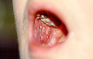
A characteristic respiratory tract complication of Granulomatosis with Polyangiitis: narrowing of the “windpipe” just below the vocal cords, a condition called “subglottic stenosis”. This narrowing, caused by inflammation and scarring, causes difficulty breathing and may, after a subacute progression, necessitate emergency tracheostomy. Pictured below are two figures that show subglottic stenosis before (left) and after (right) surgery, performed by an Ear, Nose, & Throat specialist. The surgery provided dramatic improvement in the patient’s breathing.

Lungs

A pneumonia–like syndrome, with lung “infiltrates“ can be seen on chest x–ray. Bleeding from the lungs can occur.

Kidney

Inflammation can occur in the kidney, leading to small (or rarely, large) amounts of blood and protein in the urine. This condition is called glomerulonephritis. If not treated aggressively, Granulomatosis with Polyangiitis’s involvement of the kidneys can lead to kidney failure. Renal masses can occur, but are very unusual in this disease.

The image below is from a urinalysis of a patient with kidney inflammation. When Granulomatosis with Polyangiitis is active, red blood cells will form a clump or “cast” (bracketed in white) within the tubules of inflamed kidneys. These “casts” pass through the renal system and may be viewed under the microscope in a patient’s urine.

Skin

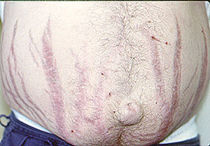
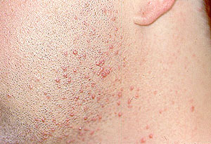
Granulomatosis with Polyangiitis can cause many kinds of skin rashes. The most common rash occurs in the form of small purple or red dots on the lower extremities (known as “palpable purpura”). Inadequate blood flow to fingers and toes can lead to Raynaud’s phenomenon (extreme sensitivity of the fingers to cold) and even infarctions of the tips of fingers and toes, with the development of gangrene.

Joints

Arthritis can occur, with joint swelling and pain.

Nerves

Peripheral nerve involvement leads to numbness, tingling, shooting pains in the extremities, and sometimes to weakness in a foot, hand, arm, or leg.

Miscellaneous

Granulomatosis with Polyangiitis involvement of nearly all organs has been described, including the meninges (the layers of protective tissue around the brain and spinal cord), the prostate gland, and the genito–urinary tract. In addition to involving specific organs, Granulomatosis with Polyangiitis also commonly results in generalized symptoms of fatigue, low–grade fever, and weight loss.

The cause of Granulomatosis with Polyangiitis is not known. Compared to diseases with obvious genetic predispositions, genetics appear to play a relatively small role in the etiology of Granulomatosis with Polyangiitis . It is very unusual for Granulomatosis with Polyangiitis to occur in two people in the same family. (It is possible, however, that less obvious genetic risk factors exist, e.g. genes that might pre-–dispose a patient to infection with an etiologic organism). For some time, an infection has been suspected of causing (or at least contributing to) Granulomatosis with Polyangiitis , but no specific infection (bacterial, viral, fungal, or other) has been identified.

How is Granulomatosis with Polyangiitis Diagnosed?

Whenever possible, it is important to confirm the diagnosis of Granulomatosis with Polyangiitis by biopsying an involved organ and finding the pathologic features of this disease under the microscope. Because many diseases may mimic Granulomatosis with Polyangiitis (and vice versa), before starting a treatment regimen it is essential to be as certain of the diagnosis as possible. We discuss some of the specific biopsy procedures used to diagnose Granulomatosis with Polyangiitis in the section of this Websie entitled What is Vasculitis: Diagnosis?.

Because Granulomatosis with Polyangiitis so often involves the upper respiratory tract (sinuses, nose, ears, and trachea [“windpipe”]) and because biopsy of these tissues is a relatively non–invasive procedure, these sites are frequently biopsied in patients suspected of Granulomatosis with Polyangiitis . Unfortunately, the yield of biopsies from these sites is rather low: probably less than 50%. Therefore, sometimes more invasive procedures are required to make the diagnosis.

Lung biopsy (either open or thoracoscopic) is often the best way of diagnosing Granulomatosis with Polyangiitis . The ample amount of tissue obtainable through these procedures usually permits confirmation of the Granulomatosis with Polyangiitis diagnosis. Similarly, although the amount of tissue obtained through a kidney biopsy is usually much smaller, the finding of certain pathologic features in the context of a patient’s overall symptoms, signs, and laboratory tests is frequently diagnostic.

Since 1982, when ANCAs (anti–neutrophil cytoplasmic antibodies) were first described, the role of these antibodies in the diagnosis of Granulomatosis with Polyangiitis has grown. ANCA testing, which involves the performance of a simple blood test, has achieved wide availability during the 1990s. This is both good and bad: use of ANCA tests has led to earlier diagnoses and more rapid institution of appropriate treatment in many cases, but has also resulted in misdiagnosis and incorrect treatment when the tests are not performed or interpreted correctly.

As their name implies, ANCAs are directed against the cytoplasm (the non-nucleus part) of white blood cells. Their precise role in the disease process remains uncertain but is a topic of considerable research interest. ANCAs come in two primary forms: 1) the C–ANCA [C stands for cytoplasmic] and, 2) the P–ANCA [P stands for perinuclear]. C–ANCAs have a particularly strong connection to Granulomatosis with Polyangiitis (up to 80% of patients — and possibly more of those with active disease — have these antibodies). When C–ANCAs are present in the blood of a patient whose symptoms or signs suggest Granulomatosis with Polyangiitis , the likelihood of the diagnosis increases considerably. In most cases, however, it is still VERY IMPORTANT to biopsy an involved organ to verify the diagnosis.

Treatment and Course of Granulomatosis with Polyangiitis

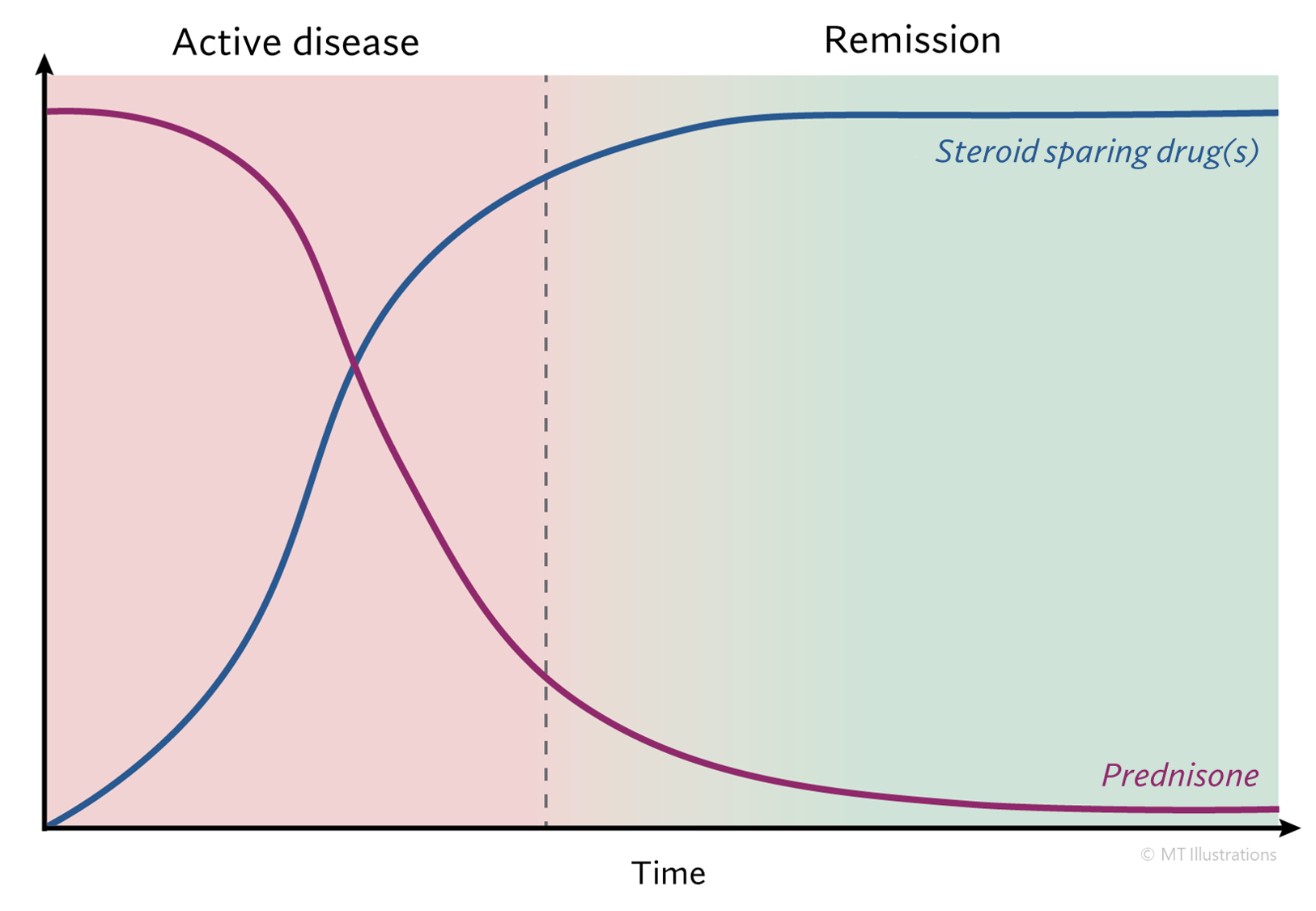
Until the 1970s, Granulomatosis with Polyangiitis was nearly always a fatal condition. The use of prednisone and other steroids helped prolong patients’ lives, but most patients eventually succumbed to the disease within a few months or years. The first use of cyclophosphamide in the late 1960s began to change the terrible prognosis of this disease. Using the combination of cyclophosphamide and prednisone, more than 90% of patients with severe disease respond to treatment, and 75% are able to achieve disease remissions. Unfortunately, Granulomatosis with Polyangiitis is a disease in which relapses frequently occur. Approximately half of all patients who achieve disease remissions eventually suffer recurrences (“flares”). Flares of Granulomatosis with Polyangiitis are usually responsive to the same treatment that induced remission, but sometimes intensification of treatment (for example, changing to a more powerful medication) is required.

During the 1990s, physicians have increasingly used the combination of methotrexate and prednisone rather than cyclophosphamide and prednisone for Granulomatosis with Polyangiitis patients who do not have immediately life-threatening disease (particularly disease that does not involve the kidneys severely), because of the frequency of severe side-effects associated with the latter regimen.

Bactrim (or Septra), a combination of two antibiotics (trimethoprim and sulfamethoxazole) may also be helpful in the treatment of Granulomatosis with Polyangiitis , particularly in patients whose disease is limited primarily to the upper respiratory tract. A large, multi-center study demonstrated that Bactrim is useful in preventing flares of Granulomatosis with Polyangiitis in the upper respiratory tract.

What’s New in Granulomatosis with Polyangiitis?

In the past few years, significant advances have been made in understanding Granulomatosis with Polyangiitis , although many important questions remain. In addition to an improved understanding of how to use the currently available medicines for Granulomatosis with Polyangiitis , it is likely that the next few years will witness the development of new medicines for this disease. Scientific breakthroughs may lead to the design of more specific modulators of the immune system that are of great benefit to patients with Granulomatosis with Polyangiitis .

In medical terms, by David Hellmann, M.D.

A discussion of Granulomatosis with Polyangiitis written in medical terms by David Hellmann, M.D. (F.A.C.P.), The Johns Hopkins Vasculitis Center, for the Rheumatology Section of the Medical Knowledge Self-Assessment Program published and copyrighted by the American College of Physicians (Edition 11, 1998). The American College of Physicians has given us permission to make this information available to patients contacting our Website

Granulomatosis with Polyangiitis is a disease involving granulomatous inflammation, necrosis and vasculitis that most frequently targets the upper respiratory tract, lower respiratory tract, and kidneys. Although Granulomatosis with Polyangiitis can begin at any age, the average age of onset is about 40 years. Other organs frequently affected by Granulomatosis with Polyangiitis granulomatosis include the eye (proptosis and double-vision from retro-orbital pseudotumor, scleritis), skin (ulcers, purpura). or peripheral nerve (mononeuritis multiplex). Granulomatosis with Polyangiitis may be limited to one site for many months or years before disseminating. Systemic symptoms (fever, fatigue, weight loss) are also common. Anemia, mild leukocytosis, and elevated Erythrocyte sedimentation rate (ESR) are nonspecific laboratory findings. Chest radiographs often show infiltrates, nodules, masses, or cavities; only hilar adenopathy is incompatible with the diagnosis of Granulomatosis with Polyangiitis. CT of the chest is more sensitive than chest radiography and can be abnormal when the chest radiograph is negative. Glomerulonephritis causes hematuria, erythrocyte casts, and proteinuria.

A novel group of autoantibodies, ANCAs, helps support the diagnosis of Granulomatosis with Polyangiitis and related forms of vasculitis and gives insight into the pathogenesis of these diseases. ANCAs are directed against enzymes cantained in primary granules of neutrophils and monocytes. Two main types of ANCAs are recognized. The C-ANCAs are directed against serine proteinase 3 and are relatively sensitive and highly specific for Granulomatosis with Polyangiitis. The P-ANCAs are directed against myeloperoxidase and other antigens and are not specific for any single form of vasculitis, but have been seen in some patients with Granulomatosis with Polyangiitis, polyarteritis nodosa, Churg-Strauss vasculitis, and some forms of pauci-immune glomerulonephritis (referred to as microscopic polyarteritis nodosa). Some patients with pulmonary-renal syndromes that may not fit the critieria for Granulomatosis with Polyangiitis are also seropositive for ANCAs. Some patients with inflammatory bowel disease, rheumatoid arthritis, or SLE may have atypical P-ANCA test results, based on the autoantibodies directed against other neutrophil constituents such as lactoferrin.

ANCAs may be not only markers for Granulomatosis with Polyangiitis and related disorders, but they may also be actors in pathogenesis. Studies show that when neutrophils are exposed to cytokines such as tumor necrosis factor, small amounts of serine proteinase and myeloperoxidase, the targets for ANCAs, are expressed on the surface of neutrophils. Adding ANCAs to these cytokine-primed neutrophils causes them to generate oxygen radicals and release enzymes capable of damaging blood vessels.

The diagnosis of Granulomatosis with Polyangiitis is established most securely by biopsy specimens showing the triad of vasculitis, granulomata, and large areas of necrosis (known as geographic necrosis) admixed with acute and chronic inflammatory cells. Only large sections of lung tissue obtained via thoracoscopic or open lung biopsy are likely to show all of the histologic features. However, more easily obtained biopsy specimens of the nose, or sinuses may show several of the changes that are highly suggestive of Granulomatosis with Polyangiitis. Such a biopsy specimen combined with a compatible clinical picture and seropositivity for C-ANCAs should suffice to secure the diagnosis. Seropositivity for C-ANCAs alone is not specific enough to establish the diagnosis of Granulomatosis with Polyangiitis.

Untreated Granulomatosis with Polyangiitis is fatal. Prednisone may slow progression of the disease but by itself is insufficient to arrest the disease. Respiratory tract disease usually progresses slowly, but renal disease can progress rapidly and therefore warrants urgent evaluation and treatment. With the traditional treatment of prednisone (initiated at 1 mg/kg daily for 1 to 2 months. then tapered) and cyclophosphamide (2mg/kg daily for at least 12 months), more than 90% of patients improve and 75% remit. However, 50% of the patients who later remit also relapse, and oral daily cyclophosphamide causes serious toxicity. Short-term toxicity includes cytopenia, infection, and hemorrhagic cystitis. Long-term use of cyclophosphamide in patients with Granulomatosis with Polyangiitis more than doubles the risk of cancer overall, increases the risk of bladder cancer 33-fold and the risk of lymphoma 11-fold. Monthly intravenous cyclophosphamide appears less toxic but also less effective. Weekly, methotrexate appears to be an effective alternative for Granulomatosis with Polyangiitis that is not immediately life-threatening, and it also appears to be beneficial in maintaining remission. The role of trimethoprim-sulfamethoxazole in treating active disease is controversial, with some finding it effective for Granulomatosis with Polyangiitis limited to the respiratory tract, and others not. In patients who have achieved remission, trimethoprim-sulfamethoxazole reduces the relapse rate.

Prednisone

Prednisone is a corticosteroid with potent anti-inflammatory effects. Corticosteroids are a cornerstone of treating most types of vasculitis, and are often used in combination with other immunosuppressive medications. Prednisone works very quickly, and is therefore used (often at high doses) at the time of initial diagnosis to bring vasculitis under control. Then, prednisone is gradually reduced (“tapered”) while another immunosuppressive drug is started for long term treatment. Over time, the “steroid-sparing” immunosuppressive drug is used to control vasculitis, and prednisone is eventually stopped.

Side Effects

Corticosteroids cause a long list of side effects, making it dangerous to use these drugs at significant doses for long term treatment. The side effects of prednisone are related to: 1) the amount of steroid a patient takes in his/her daily dose, and 2) the length of time the patient remains on the medication. We emphasize that not all side effects occur in all patients.

Despite the numerous potential side effects of corticosteroids listed below, their introduction into patient care more than 50 years ago revolutionized the treatment of many diseases, including vasculitis. When used properly, these drugs save lives and avert threats to the function of important organs.

Common Side Effects of Steroids:

  • Increased Susceptibility to Infections
  • Weight Gain
  • Glucose Intolerance
  • Hypertension
  • Bone Thinning
  • Avascular Necrosis of bone
  • Easy Bruising
  • Abdominal Striae
  • Hirsutism
  • Acne
  • Mood Swings/Insomnia
  • Cataracts

Increased Susceptibility to Infections

Patients are at increased risk for many types of infections, from minor fungal infections in the mouth (“thrush”, caused by Candida) to life–threatening infections such as Pneumocystis carinii pneumonia. The higher the steroid dose and the longer the duration of therapy, the greater the risk of infection. The risk is also increased when patients receive combinations of immunosuppressive medications, such as cyclophosphamide (cytoxan) and prednisone. The risk of some infections can be greatly reduced by taking specific types of antibiotics prophylactically (such as Bactrim).

Pictured below is woman under treatment with prednisone and methotrexate for vasculitis and a concurrent neurologic condition (myasthenia gravis) developed painful vesicles in her mouth. The vesicles were confirmed by culture to be caused by reactivation of a Herpes simplex infection, and responded to treatment with acyclovir.

Weight Gain

Weight gain is usually the most dreaded side–effects of steroids, incurred to some degree by nearly all patients who take them. The amount of weight gain varies from individual to individual. In addition to causing weight gain, prednisone leads to a redistribution of body fat to places that are undesirable, particularly the face, back of the neck, and abdomen. Pictured below is an example of redistribution of body fat to the back of the neck. Accumulation of fat in this area is sometimes referred to as a “buffalo hump”.

Another example of this “redistribution” is pictured below. Supraclavical “fat pads” are collections of fat at the base of the neck, just above the collarbones, which are common in patients on steroids. They sometimes cause concern among patients if mistaken for lymph nodes or other causes for worry, but will gradually subside as the prednisone dose is tapered to below 10 milligrams/day.

In addition to this redistribution of fat, many patients undergo loss of muscle strength (muscle atrophy) while taking steroids. Regular physical exercise is key to avoiding this type of deconditioning that often occurs with prednisone treatment.

Glucose Intolerance

High blood sugar, or steroid–induced diabetes. Patients who are “pre-diabetic” can develop diabetes and the need for insulin while taking steroids. This usually resolves when the steroids are decreased or discontinued, but can be worsened by weight gain.

Hypertension

High blood pressure. This usually improves as the corticosteroid dose is reduced.

Bone Thinning (Osteoporosis)

Prednisone may cause thinning of the bones even in people who are not usually at high risk for osteoporosis (for example: males, young people). In people susceptible to osteoporosis, prednisone may accelerate the process of bone loss. Fortunately, in the past few years, excellent treatments and preventive measures have become available for osteoporosis. All patients on prednisone for prolonged periods are candidates for these medicines. Patients should be aware of their daily intake of calcium and Vitamin D while on steroids. Bone density measurement is commonly done using DEXA scans.

Avascular Necrosis of Bone

For reasons that are not known, high dose prednisone (for example, greater than 20 milligrams a day) predisposes some patients to joint damage, most often of the hips. In avascular necrosis (or osteonecrosis, meaning “bone death”) of the hip, the part of the leg bone that inserts into the pelvis dies, resulting in pain with weight–bearing and some loss of joint function. Many patients with avascular necrosis require joint replacements.

Easy Bruising

Prednisone also causes “thin skin”. Patients on moderate to high doses of prednisone often notice that they bruise easily, even with only slight trauma. Pictured below is a patient with giant cell arteritis who suffered a skin laceration after she struck her leg against a chair.

Abdominal Striae

Abdominal striae (“stripes”), as pictured below, frequently occur in patients who take high doses of steroids for long periods of time.

Hirsutism

Hirsutism is excessive growth of body hair. Patients vary in the degree to which this side effect of steroids occurs. Although some patients experience minimal hirsutism, the patient depicted below developed this side effect after taking 10 milligrams of prednisone for a few months.

Acne

High dose prednisone predisposes some patients to acne, especially facial acne, as pictured below. The facial acne developed after several weeks of high steroid doses.

Mood Swings/Insomnia

Many patients find it difficult to fall asleep when taking high doses of steroids. Many also find that they are more irritable or anxious than usual. Steroids sometimes even induce depression or psychosis, which usually improves when the drug is decreased or discontinued.

Cataracts

Long–term steroid use may lead to cataract development in the eyes, which frequently require surgical removal.

Microscopic Polyangiitis

  • First Description
  • Who gets Microscopic Polyangiitis (the “typical” patients)?
  • Classic symptoms of Microscopic Polyangiitis
  • Forms of vasculitis similar to Microscopic Polyangiitis
  • What causes Microscopic Polyangiitis?
  • How is Microscopic Polyangiitis diagnosed?
  • Treatment and Course of Microscopic Polyangiitis

First Description

The first description of a patient with the illness now known as microscopic polyangiitis (MPA) appeared in the European literature in the 1920s. The concept of this disease as a condition that is separate from polyarteritis nodosa (PAN) and other forms of vasculitis did not begin to take root in medical thinking, however, until the late 1940s. Even today, some confusing terms for MPA (e.g., “microscopic poly arteritis nodosa ” rather than “microscopic poly angiitis ”) persist in the medical literature. Confusion regarding the proper nomenclature of this disease led to references to “microscopic polyarteritis nodosa” and “hypersensitivity vasculitis” for many years. In 1994, The Chapel Hill Consensus Conference recognized MPA as its own entity, distinguishing it in a classification scheme clearly from PAN, granulomatosis with polyangiitis (GPA, formerly Wegener’s), cutaneous leukocytoclastic angiitis (CLA), and other diseases with which MPA has been confused with through the years.

Much of the explanation for the difficulty in separating MPA from other forms of vasculitis has stemmed from the numerous areas of overlap of MPA with other diseases. MPA, PAN, GPA, and CLA  and other disorders all share a variety of features but possess sufficient differences as to justify separate classifications.

Who gets Microscopic Polyangiitis? A typical patient

MPA can affect individuals from all ethnic backgrounds and any age group. In the United States, the typical MPA patient is a middle-aged white male or female, but many exceptions to this exist. The disease may occur in people of all ages, both genders, and all ethnic backgrounds.

Classic symptoms of Microscopic Polyangiitis

Many signs and symptoms are associated with MPA. This disease can affect many of the body’s organ systems including (but not limited to) the kidneys, nervous system (particularly the peripheral nerves, as opposed to the brain or spinal cord), skin, and lungs. In addition, generalized symptoms such as fever and weight loss are very common.

The FIVE most common clinical manifestations of MPA are:

  1. Kidney inflammation (~ 80% of patients).
  2. Weight loss (> 70%).
  3. Skin lesions (> 60%).
  4. Nerve damage (60%).
  5. Fevers (55%).

Kidney Inflammation

Inflammation in the kidneys, known as glomerulonephritis, causes blood and protein loss through the urine. This process can occur either slowly or very rapidly in the course of the disease. Patients with kidney inflammation may experience fatigue, shortness of breath, and swelling of the legs.

The image below is from a urinalysis of a patient with kidney inflammation. When MPA is active, red blood cells will form a clump or “cast” (bracketed in white) within the tubules of inflamed kidneys. These “casts” pass through the renal system and may be viewed under the microscope in a patient’s urine.

Constitutional Symptoms

Weight loss, fevers, fatigue, and malaise are part of a collection of complaints regarded as “constitutional” symptoms. Constitutional complaints are a common finding in patients with MPA, because the disorder is a systemic disease confining itself generally not to one specific organ system but rather broadly affecting a patient’s “constitution”.

Skin lesions

Skin lesions in MPA, as in other forms of vasculitis that involve the skin, can erupt on various areas of the body. The lesions tend to favor the “dependent” areas of the body, specifically the feet, lower legs and, in bed-ridden patients, the buttocks. The skin findings of cutaneous MPA include purplish bumps and spots pictured below (palpable purpura).

These areas range in size from several millimeters in diameter to coalescent lesions that are even larger. Skin findings in MPA may also include small flesh-colored bumps (papules); small-to-medium sized blisters (vesiculobullous lesions); or as small areas of bleeding under the nails that look like splinters (pictured below), hence the name splinter hemorrhages.

Peripheral nervous system

Damage to peripheral nerves (i.e., nerves to the hands and feet, arms and legs) results from inflammation of the blood vessels that supply the nerves with nutrients. Inflammation in these blood vessels deprives the nerves of their nutrients, leading to nerve infarction (tissue death). Multiple nerve involvement that is characteristic of vasculitis is known as “mononeuritis multiplex”. This condition is frequently associated with wrist or foot drop: the inability to extend the hand “backwards” at the wrist or to flex the foot upward toward the head at the ankle joint. If the condition is caused by nerve deterioration associated with vasculitis, unfortunately, surgery is not a treatment option due to the nerve infarcton (tissue death).

Neurologic symptoms resulting from peripheral nerve damage may also include numbness or tingling in the arm, hand, leg, or foot. Over time, muscle wasting (pictured below) that is secondary to the nerve damage may result from damage caused by vasculitis.

Pictured:

The hand on the left (the patient’s right hand) is normal, displaying normal muscle bulk of the areas between the fingers.  In contrast, the hand on the right (the patient’s left) shows wasting of the muscle in the web space between the thumb and first finger, leading to a hollowed-out, bowl-like appearance of that area.  The consequence of this muscle wasting is that the patient is unable to grasp objects between his thumb and fingers (i.e., has a weak pinch) and his hand grip is weak.

Lungs

Lung involvement can be a dramatic and life-threatening manifestation of MPA. When lung disease takes the form alveolar hemorrhage – bleeding from the small capillaries that are in contact with the lungs’ microscopic air sacs – the condition may quickly pose a threat to the patient’s respiratory status (and therefore to the patient’s life). Alveolar hemorrhage (pictured below), which is frequently heralded by the coughing up of blood, occurs in approximately 12% of patients with MPA .

Another common lung manifestation of MPA is the development of non-specific inflammatory infiltrates, identifiable on chext x-rays or computed tomography (CT scans) of the lung.

Eyes, Muscles, and Joints

Organs that also merit mention in discussions of MPA include the eyes, muscles, and joints. Intermittent irritation of the eye (resembling “pinkeye”) that is caused by either conjunctivitis or episcleritis may be an early disease manifestation or a sign of a disease flare. Occasionally other types of inflammation (e.g., uveitis) are also observed in MPA. Muscle or joint pains (known to clinicians as “myalgias” or “arthralgias”, respectively) are common complaints in MPA, generally accompanying the types of constitutional symptoms mentioned above. Arthritis (inflammation of the joints accompanied by swelling) can also be observed in MPA. Joint complaints in MPA and related forms of vasculitis tend to migrate from one joint to another – one day involving the left ankle, the next day the right wrist, the third day a shoulder, for example.

Forms of vasculitis similar to Microscopic Polyangiitis

The similarities and differences between MPA, GPA, and PAN are highlighted in the table below.

MPA GPA PAN
BLOOD VESSEL SIZE Small to Medium Small to Medium Medium
BLOOD VESSEL TYPE Arterioles to venules, And sometimes Arteries and veins Arterioles to venules, And sometimes Arteries and veins Muscular Arteries
GRANULOMATOUS INFLAMMATION NO YES NO
LUNG SYMPTOMS YES1 YES1 NO
GLOMERULONEPHRITIS YES YES NO
RENAL HYPERTENSION NO NO YES
MONONEURITIS MULTIPLEX COMMON OCCASIONAL COMMON
SKIN LESIONS YES2 YES2 YES2
GI SYMPTOMS NO NO YES3
EYE SYMPTOMS YES4 YES4 NO
ANCA-POSITIVITY 75% 65-90% NO
CONSTITUTIONALSYMPTOMS YES5 YES5 YES5
NECROTIZING TISSUE YES YES YES
MICROANEURYSMS RARELY RARELY TYPICAL

1 Pulmonary capillaritis in MPA and nodules or cavitary lesions in WG

2MPA can have small blood vessel skin lesions as mentioned above, similar to GPA or medium blood vessel lesions similar to PAN (livedo reticularis, nodules, ulcers, and digital gangrene)

3Stomach pain after meals

4MPA eye complications are typically milder than those of GPA, but serious

ocular problems including necrotizing scleritis can occur

5Constitutional symptoms include weight loss, fevers, joint and muscle aches, and malaise.

What Causes Microscopic Polyangiitis?

The cause of MPA is not known. However, enough is known about a few types of vasculitides that allow us to describe in general terms how MPA affects the body. MPA is clearly a disorder that is mediated by the immune system; the precise events leading to the immune system dysfunction (hyperactivity), however, remain unclear. Many elements of the immune system are involved in this process: neutrophils, macrophages, T and B lymphocytes, antibodies, and many, many others.

Because MPA is often associated with anti-neutrophil cytoplasmic antibodies (ANCA), antibodies directed against certain constituents of white blood cells (WBCs), the disease is often termed an “ANCA-associated vasculitis”, or AAV. ANCA, discovered in 1982, act against certain specific (and naturally occurring) enzymes in the body residing within the neutrophils and the macrophages, all of which are members of the WBC family. The result of the interactions of ANCA with their target proteins is an increase in the destruction of WBCs at the sites of disease and the release of white blood cell enzymes within blood vessel walls, causing the damage to blood vessels. In MPA, the ANCA are directed generally against to specific proteins: myeloperoxidase (MPO) and proteinase 3 (PR3).

How is Microscopic Polyangiitis diagnosed?

Blood is taken to detect any ANCA levels, if MPA is suspected. In addition, an erythrocyte sedimentation rate (ESR or “sed rate”) and C-reactive protein (CRP) are usually ordered. Both of these tests are elevated in many different types of inflammation and are not specific to MPA or any particular disease. The ESR and CRP, known as “acute phase reactants”, are often sensitive indicators of the presence of active disease. In and of themselves, however, elevations in acute phase reactants are not sufficient to justify additional treatment.

A carefully analyzed urine specimen should be obtained at the initial visit (and every follow-up visit!) to maintain vigilance for either the development or the progression of kidney involvement.

A computed tomography (CT) scan of the chest may also be performed to detect the presence of lung involvement. A tissue biopsy may be needed to make the diagnosis of MPA, and is taken from an organ that seems to be involved at the time. Sometimes an electromyography/nerve conduction (EMG/NCV) study may need to be done to identify a site for biopsy or to detect findings consistent with a mononeuritis multiplex (see classic symptoms section above). Tissues that might be biopsied are kidney, skin, nerve, muscle, and lung.

Pictured: a biopsy of the gastrocnemius muscle, performed in a 69 year–old man with microscopic polyangiitis. A blood vessel within the muscle shows an intense inflammatory infiltrate with destruction of the blood vessel wall, confirming the diagnosis of vasculitis.

Treatment and Course of Microscopic Polyangiitis

A steroid (usually prednisone) in combination with a cyclophosphamide (CYC) or rituximab is typically the first combination of medications to be prescribed.  After control of the disease – usually around 4 – 6 months of treatment maintenance therapy will be used to keep the disease in remission. This will vary between patients. Prednisone may be discontinued after approximately 6 months.

Research Information

Research Information

Research and clinical trials are essential for the advance of medicine. The effectiveness of each new breakthrough can only be assessed by putting them into practice with individuals affected by the disease.

For those who care about vasculitis and discovering new ways to identify, diagnose, and treat these diseases, there are many ways to support the advancement of research. Volunteer participation in clinical trials is one way to help progress knowledge about the various types of vasculitis. As a volunteer, your participation can help potentially new, successful treatments become available to others who understand living with vasculitis as you do. For certain non-medication based studies, a volunteer may be any individual who is willing to participate. Volunteers can be spouses, friends and family members who wish to make a difference in vasculitis research.

Open Research Studies

Clinical Epidemiology and serological/plasma and genetic factors in systemic vasculitis

Purpose of Study:

This research is being done to gather information about patients with certain vasculitis diagnoses. The Johns Hopkins Vasculitis Center is building a comprehensive database of information concerning vasculitis patients as part of its mission to facilitate research.

Recruiting patients:

  • Diagnosed with vasculitis
  • Patients of the Johns Hopkins Vasculitis Center

Coordinator/Contact: Hannah Smith
Email: hsmith97@jh.edu
Phone number: 410-550-0122
Principal Investigator: Duvuru Geetha, M.D.
IRB#: IRB00355688

A Randomized, Double-blind, Placebo-controlled Phase 4 Clinical Trial to Evaluate the Long-term Safety and Efficacy of Avacopan in Subjects With Antineutrophil Cytoplasmic Antibody (ANCA)-associated Vasculitis

Purpose of the Study:

This study is to make sure the medicine called avacopan is safe to use for a long time. It will check for liver problems and serious allergic reactions. It will also track the medicine keeps the illness in remission, kidney function, and how the treatment affects daily life.

Recruiting patients:

  • Newly diagnosed or relapse of GPA or MPA
  • Age 18 years or older
  • Positive test for anti-PR3 or anti-MPO (current or historic) antibodies
  • Patients of the Johns Hopkins Vasculitis Center

Coordinator/Contact: Hannah Smith
Email: hsmith97@jh.edu
Phone number: 410-550-0122
Principal Investigator: Duvuru Geetha, M.D.
IRB#: IRB00403470

Frequently Asked Questions about Clinical Trials

From the definition of a clinical trial to information and questions that are good to ask your doctor before participating in a trial, this section address many of the common questions that arise when deciding to join in supporting vasculitis research.

What is a clinical trial?

A clinical trial is a type of research study. Clinical trials test a new treatment and compare it to the available treatment (the usual way doctors treat a certain health condition or disease). For example, a clinical trial might study how well a new medicine helps people with cancer or if certain foods help people stay healthy. The Food and Drug Administration requires clinical trials before a new medication can be approved. Sometimes it is necessary to compare an experimental treatment with a placebo (inactive treatment) when no standard treatment exists.

FDA Consumer magazine published this article “Inside Clinical Trials Testing Medical Products in People” which may assist in understand the purpose, process and procedure of clinical trials from the Food and Drug Administrative’s perspective. 

Why should I take part in a clinical trial?

Because the trials are investigations designed to learn more about a specific disease or treatment, personal benefit cannot be guaranteed. The benefits of taking part in a clinical trial depend on the study you join. The benefits also depend on your assigned study group. Here are some possible benefits you might get from taking part in a clinical trial. You may:

  • get free health exams;
  • learn more about your health;
  • take a more active role in your own health care;
  • have your health watched closely;
  • receive some medications at no cost to you; and help answer research questions that may mean better health for people in the future.

What are the risks of taking part in a clinical trial?

There are risks to you when you take part in a clinical trial. The study doctors and coordinators will watch you carefully for any changes in your health. You are always free to leave the study. The risks will vary depending on the kind of trial you join. Here are some possible risks.

  • You may have side effects (health problems) from taking a new medicine or getting a new procedure that is being tested. There may be side effects that are unexpected. Usually you will need to give blood samples. Some people find that process uncomfortable.
  • The visits for the clinical trial may be frequent and time consuming.

The therapy you receive may not be effective or you may be assigned to a placebo group

How do I know if I have been given all the information I need about taking part in a clinical trial?

When beginning any study the doctor, or investigator, must ask approval from an Institutional Review Board (IRB). The IRB is a committee of doctors and other medical personnel that have no ties to the study. The IRB makes sure the study is as safe as possible and that the “informed consent” explains all of the important information to the patient.

Before people join a clinical trial, they go through something called the “informed consent process”. This process means that you are given written information that tells you about the purpose of the study; risks and benefits of being in the study; and what will happen to you in the study. You will be given an informed consent form, which you will need to read over very carefully. Take the form home and share it with family members, friends, and your primary care doctor. Once you have read the form, ask questions about words or procedures that you don’t understand.

Another part of the informed consent process is that you can ask questions about the study at any time. It is your right to have all the information you need to make your decision about whether or not to take part in a clinical trial. Don’t let anyone pressure you into taking part in a clinical trial. The choice is yours.

What if I decide that I don’t want to be a part of the study, even though the study has already started?

That’s okay. You can change your mind and leave the study at any time. Remember that being a part of a clinical trial is always your choice. Your relationship with your doctor will not change because you decide to leave the study. Your care will not be affected in any way.

Who takes part in clinical trials?

Many different people take part in clinical trials. People who take part in clinical trials are volunteers who meet eligibility criteria for the study. Eligibility criteria are requirements that someone must meet to be a part of the study. Some examples of eligibility criteria are having a certain disease such as Wegener’s Granulomatosis (WG); not showing improvement on standard WG medications; being a certain age; or being in good health. These criteria help make sure that the study answers the right research question.

Is it safe to participate in a clinical trial if trying to conceive a child or pregnant?

Check with your doctor or the study coordinator to find out if it is safe to participate in a particular study if you are trying to conceive, if you are pregnant or postpartum. If the trial is assessing medications, there is often concern about how medicines used in a study could affect a pregnancy regardless of being male or female. The informed consent form should tell you if any of the medicines in the study could affect a pregnancy.

For women, if you are postpartum and breast feeding, check with your doctor to make sure it is okay to breast feed your baby while you are taking part in a clinical trial. Don’t be afraid to ask questions about safety.

How can I find out about clinical trials or other research studies at the Johns Hopkins Vasculitis Center ?

If you would like more information about one of our research activities or if you would like to be a participant, please contact the Johns Hopkins Vasculitis Center.

Eosinophilic Granulomatosis with Polyangiitis, formerly Churg-Strauss Syndrome (EGPA)

  • First Description
  • Who gets EGPA (the “typical” patients)?
  • Classic symptoms of EGPA
  • What causes EGPA?
  • How is EGPA diagnosed?
  • Treatment and Course of EGPA

First Description

Eosinophilic granulomatosis with polyangiitis (EGP), formerly known as the Churg-Strauss Syndrome , is a systemic vasculitis. This disease was first described in 1951 by Dr. Jacob Churg and Dr. Lotte Strauss as a syndrome consisting of “asthma, eosinophilia [an excessive number of eosinophils in the blood], fever, and accompanying vasculitis of various organ systems”. EGPA shares many of the clinical and pathological features of polyarteritis nodosa (“PAN”, another type of vasculitis) and granulomatosis with polyangiitis (GPA). However, Drs. Churg and Strauss discovered that the presence of granulomas as well as the abundance of eosinophils distinguished this disease from PAN and GPA.

Who gets EGPA (the “typical” patient)?

The typical patient with EGPA is a middle aged individual with a history of new-onset or newly-worsened asthma. The distribution of the disease among males and females is approximately equal.

Classic symptoms and signs of EGPA

Asthma is one of the cardinal features of EGPA. Asthma symptoms may begin long before the onset of vasculitis – e.g., many years before any other symptoms of EGPA arise, and long before the diagnosis of EGPA is made. Other early symptoms/signs include nasal polyps and allergic rhinitis.

The next phase of the disease is often marked by eosinophilia, the finding of an excessive number of eosinophils in the blood or in tissues. An eosinophil is one subtypes of white blood cell. Normally, eosinophils comprise 5% or less of the total white blood cell count. In EGPA, the percentage of eosinophils may reach as high as 60%. In the picture below, the eosinophils are shown by the dark pink stain.

The third phase of the illness is a vasculitis, which involves the skin, lungs, nerves, kidneys, and other organs. Particular mention should be made of the frequent devastating involvement of the nerves (called mononeuritis multiplex), which produces severe tingling, numbess, shooting pains, and severe muscle wasting/power loss in the hands or feet. The list below contains the organs commonly involved by EGPA and the specific disease manifestation(s) in each organ.

  • Nose
  • Lung
  • Skin
  • Kidney
  • Gastrointestinal
  • Heart
  • Nerve

Nose

  • Sinusitis, including allergic rhinitis
  • Nasal polyps

Lung

  • Pulmonary infiltrates (only one-third of all patients)
  • Bleeding into the lungs (occasionally)
  • Diffuse interstitial lung disease (rarely)

Skin

  • Rashes
  • Palpable purpura
  • Nodules (above or below the skin), often at sites of pressure, such as the elbows

Kidney

  • Glomerulonephritis (inflammation in the small units of the kidney that filter blood)
  • Hypertension

Gastrointestinal

  • Lesions (vasculitic) are occasionally found in the GI tract
  • Granuloma sometimes found in spleen

Heart

  • Vasculitis lesions in heart, can lead to congestive heart failure or a heart attack

Nerve

  • Peripheral nerve involvement including pain, numbness, or tingling in extremities (neuropathy/mononeuritis multiplex)

What causes EGPA?

The cause of EGPA is unknown but is probably multi-factorial. Genetics may play a small role in the disease, but EGPA is almost never seen in two members of the same family. Environmental factors such as exposure to industrial solvents may play a role in susceptibility to this disease, but this is largely speculative. Infections may be the inciting event(s), but to date there is no definitive evidence of this.

How is EGPA Diagnosed?

Among all of the vasculitides, asthma is a distinctive feature of EGPA alone. However, not all patients with asthma have vasculitis – only a tiny minority do, in fact. It is the specific combination of symptoms and signs, the pattern of organ involvement, and the presence of certain abnormal blood tests (eosinophilia, in particular) that help the doctor make the diagnosis. In addition to a detailed history and physical examination, blood tests, chest X-rays and other types of imaging studies, nerve conduction tests, and tissue biopsies (e.g., of lung, skin, or nerve) may be performed to help diagnose EGPA.

The following features are consistent with a diagnosis of GPA:

  1. asthma
  2. eosinophilia [>10% on differential WBC count]
  3. mononeuropathy
  4. transient pulmonary infiltrates on chest X-rays
  5. paranasal sinus abnormalities
  6. biopsy containing a blood vessel with extravascular eosinophils.

Treatment and Course of EGPA

EGPA usually responds to prednisone. Initially, high doses of oral prednisone are used in an attempt to get the disease into remission as quickly as possibly (e.g., using oral prednisone 40-60 mg/day). After the first month or so, this high dose of prednisone is gradually tapered down over the ensuing months. Other immunosuppressive drugs, such as azathioprine, cellcept, methotrexate, cyclophosphamide, or rituximab may be used in addition to prednisone. High doses of intravenous steroids (usually methylprednisolone) maybe useful for those patients with severe disease or for those who are unresponsive to the combination of oral prednisone used with other immunosuppressive medications.

Prior to the advent of prednisone, EGPA was often a fatal disease. The majority of patients died from rampant, uncontrolled disease. With present therapy, constitutional symptoms begin to resolve quite quickly, with gradual improvement in cardiac and renal function, as well as improvement in the pain that results from peripheral nerve involvement. The course of therapy can last for 1 to 2 years, although the length and type of treatment depend on the severity of disease and the organs involved. The patient’s response to treatment and the continuation of disease control during lowering of the prednisone dose are the primary determinants of how long therapy is continued. Laboratory monitoring of blood tests is very helpful in gauging the activity of disease. Some of the most useful laboratory tests are the erythrocyte sedimentation rate (ESR) and the eosinophil count.

Vasculitis Treatments

Vasculitis Treatments

While there are many different forms of vasculitis, all share the feature of organ damage caused by the immune system. Therefore, treatments for vasculitis are medications that suppress parts of the immune system (immunosuppressant drugs). The earliest treatment strategies developed for vasculitis involved very broadly-acting immunosuppressive drugs that lower immune system function in a relatively non-specific way. Today, vasculitis patients benefit from more sophisticated modern therapies that target specific portions of the immune system.

General Principles

We use a variety of medications, sometimes in combinations, to treat different forms of vasculitis. Yet a similar general approach is used across these diseases. Active vasculitis causes damage to the organs affected by the disease. We act quickly to stop the inflammation that is present in active disease. When successful, a patient then enters a state of disease remission. During remission, all signs of active inflammation are gone, but there may still be symptoms due to damage in organs where inflammation was previously present.

When disease is in an active state we typically use steroids (prednisone) to bring the disease under control quickly. The amount and type of steroids used depends on the severity of the inflammation present. Generally, steroids are used on a short-term basis to bring the disease under initial control (remission). We avoid long-term use of steroids because of their numerous and serious side effects. Some mild forms of vasculitis do not require any prednisone at all.

Along with the initiation of steroids, we typically start a “steroid-sparing” drug that will be used on a longer-term basis. These steroid-sparing drugs provide the antiinflammatory effect that controls vasculitis, while avoiding the side effects of steroids. Some steroid-sparing drugs are “biologic” medications – drugs that are made from cells, rather than chemicals synthesized in a reaction or extracted from a natural source. Examples of biologic treatments include insulin, vaccines, and blood products. In rheumatology, many biologics are antibodies that have been designed to target specific parts of the immune system. These drugs provide greater specificity than older drugs, therefore helping to reduce side effects. In addition to immunosuppressive drugs, we use some other treatments that are immunomodulatory in function – they have an impact on the immune system, but do not lower immune function in such a way that the risk of infection is increased. Examples of immunomodulatory drugs include colchicine and IVIG.

The first step in treating active vasculitis is to bring the disease into a state of remission. Prednisone impacts the immune system very quickly, and does the bulk of the work to reduce inflammation acutely. Most steroid-sparing drugs take longer to have an effect. After active disease has been adequately treated, our next goal is to reduce and then eliminate steroids, and use the steroid-sparing drug alone to maintain disease remission. Sometimes we are able to switch to milder immunosuppressants to maintain disease remission for longer timeframes. Occasionally, we are forced to maintain the use of prednisone for a longer time, but we always work to minimize the amount of prednisone used in these cases.

Once patients are successfully into remission, we continue using maintenance immunosuppression to keep the disease in a quiet state. The overall duration of therapy depends on many factors, including: the specific type of vasculitis present and its propensity to relapse, the severity and type of organ damage that was present at onset, the extent to which patients tolerate immunosuppression, other medical diagnoses that may be present, and other factors. Most forms of vasculitis have the ability to return to an active state at some point in the future, which is referred to as a relapse or flare of disease. In general, the symptoms that occur during relapses are less severe than those that were present at the beginning of a patient’s illness before immunosuppression had been initiated.

Below you will find information on the specific drugs that we utilize in the management of vasculitis.

Vasculitis medications:

  • Prednisone
  • Avacopan
  • Apremilast
  • Azathioprine
  • Colchicine
  • Cyclophosphamide
  • Dapsone
  • Intravenous or Subcutaneous Immunoglobulin (IVIG/SCIG)
  • Leflunomide
  • Mepolizumab
  • Methotrexate
  • Mycophenolate
  • Rituximab
  • Sarilumab
  • TNF inhibitors
  • Tocilizumab

All information contained within the Johns Hopkins Vasculitis Center website is intended for educational purposes only. Visitors are encouraged to consult other sources and confirm the information contained within this site. Consumers should never disregard medical advice or delay in seeking it because of something they may have read on this website.

  • Go to page 1
  • Go to page 2
  • Go to page 3
  • Interim pages omitted …
  • Go to page 6
  • Go to Next Page »

Primary Sidebar


donate to the Johns Hopkins Vasculitis Center

All information contained within the Johns Hopkins Vasculitis Center website is intended for educational purposes only. Visitors are encouraged to consult other sources and confirm the information contained within this site. Consumers should never disregard medical advice or delay in seeking it because of something they may have read on this website.

Footer

Johns Hopkins Rheumatology

  • Johns Hopkins Rheumatology
  • Johns Hopkins Arthritis Center
  • Johns Hopkins Lupus Center
  • Johns Hopkins Lyme Disease Research Center
  • Johns Hopkins Myositis Center
  • Johns Hopkins Scleroderma Center
  • Johns Hopkins Sjögren’s Syndrome Center

Connect With Us

  • Facebook
  • Twitter
  • YouTube
U.S. News and World Report Rankings Badge

Johns Hopkins Medicine Johns Hopkins Medicine logo
Language Assistance Available:
  • Español
  • American Sign Language
  • עִברִי
  • אידיש
  • አማርኛ
  • 繁體中文
  • Français
  • Tagalog
  • Русский
  • Português
  • Italiano
  • Tiếng Việt
  • Ɓàsɔ́ɔ̀-wùɖù-po-nyɔ̀
  • Igbo asusu
  • èdè Yorùbá
  • বাংলা
  • 日本語
  • 한국어
  • Kreyòl Ayisyen
  • العربية
  • Deutsch
  • Polski
  • Ελληνικά
  • ગુજરાતી
  • ภาษาไทย
  • اُردُو
  • فارسی
  • हिंदी
  • Deitsch
  • ខ្មែរ
  • မြန်မာ
Contact & Privacy Information
Price Transparency
Notice of Privacy Practices Privacy Statement
Terms & Conditions of Use Non-Discrimination Notice
Manage Cookie Preferences
Copyright © 2025 Johns Hopkins Rheumatology