• Skip to primary navigation
  • Skip to main content
  • Skip to primary sidebar
  • Skip to footer

Johns Hopkins Vasculitis Center

Show Search
Hide Search
  • About Our Center
    • Appointments and Directions
    • Meet Our Team
    • Support Our Center
  • What is Vasculitis?
    • Types of Vasculitis
    • Causes of Vasculitis
    • Symptoms of Vasculitis
    • Diagnosing Vasculitis
  • Vasculitis Treatments
    • Prednisone
    • Avacopan (Tavneos®)
    • Apremilast (Otezla®)
    • Azathioprine
    • Colchicine
    • Cyclophosphamide (Cytoxan)
    • Dapsone
    • Supplemental Immunoglobulin (IVIG/SCIG)
    • Leflunomide
    • Mepolizumab (Nucala®)
    • Methotrexate (MTX)
    • Mycophenolate
    • Rituximab
    • Sarilumab (Kevzara®)
    • TNF Inhibitors
    • Tocilizumab (Actemra®)
  • Vasculitis Research
  • Resources
Home / Search for "search.html/treatments/methotrexate.html/whatis/diagnosis.html"

Search Results for: search.html/treatments/methotrexate.html/whatis/diagnosis.html

Linda’s Loop

contributed by Brenda Shilling

I have always known that chronic and life-threatening diseases can have a devastating effect on people’s lives. I’ve seen it happen to friends’ families and heard of it happening to friends of friends. But until 2002, the realities of such challenges were quite remote to me, as I had never felt a loved one struggle with a serious condition.

Linda Gray and her twin sister, Brenda Shilling.

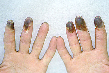
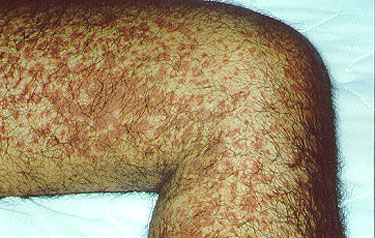
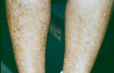
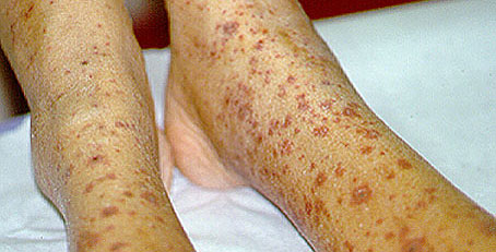
In the spring of 2002, my identical twin, Linda, was diagnosed with neuropsychiatry lupus and central nervous system vasculitis. It was a scary time for all of us, particularly for Linda and her family. Like many lupus patients, Linda had been ill often and had many problems in the years leading up to her diagnosis. We were extremely thankful that The Johns Hopkins Vasculitis Center quickly identified what was wrong and started Linda on a treatment program.

In the months that followed, I watched my sister experience a multitude of emotions, including devastation over her diagnosis, fear for her future, and relief that she had finally found help. Although I know that not all lupus patients fare well, I learned that the treatment of lupus has come a long way over the years and I prayed that Linda would benefit from these advances. Her treatment was extremely challenging. However, during the hard realities of chemotherapy and steroids on Linda’s body, her spirit was amazing.

Despite knowing that family is extremely important during times like this and that frequent calls, letters, and visits to Linda were supportive, I slowly began to feel helpless. This was an unexpected emotion. I wanted to do more. I needed to do more. My sister was going through the most difficult time of her life, and I had do something more.

One morning in the late spring of 2003 I considered coordinating a bicycle ride to raise funds for The Johns Hopkins Vasculitis Center. Our sister, Liz, thought it was wonderful idea and wanted to help. We were driven by love for our sister and that was all the motivation we needed!

Linda Gray and her sister, Liz Adams.

We decided on a 50-mile ride – short enough to manage but long enough to sound good! We then mailed over 200 letters to friends and family asking for a financial gift to The Johns Hopkins Vasculitis Center in honor of our sister and others with her disease. We also distributed the letter to social and religious groups in which we are involved. Four of our friends asked if they could join us on the bike ride in a generous gesture of support. We even had t-shirts made!

Linda's Loop Participants

Participants in Linda’s Loop:

Left to Right: (Front Row, kneeling) Georgann Pattillo, Jan Rowe, Bill Schilling ; (Middle Row) Kevin Adams, Linda Moore, Liz Adams, Sue Schilling, Brenda Schilling, Betty Lamey, Patrick Pattillo, Leroy Lamey ; (Back Row) Chandler Burroughs, Steven Rowe, and Bill Schilling, Sr.

The night before the ride, we finished packing up drinks, snacks, and lunches and then headed home to get some sleep. Liz told me that she didn’t sleep a wink that night – neither did I! We were too excited.

The day finally arrived and everything went so well. It was such a wonderful day that I simply didn’t want it to end! Our family and friends came out to cheer us along. It felt great to have them there. But wow! The miles were more difficult than I expected. Although three riders finished far ahead, the remaining three of us dragged ourselves over the finish line some time later. We were quite the motley crew!

Hot, sweaty, hungry, but happy!

Response to our effort was overwhelming. My husband Bill teases that this was the only time I actually picked up the mail! Liz and I were moved by all the contributions. Some were from people who don’t know Linda but had read about the ride in the local paper. When we started planning we weren’t sure how much money we could raise. It was wonderful to have received just over $8,600 in contributions to vasculitis research!

Linda told me how much love she felt because of what we did for The Johns Hopkins Vasculitis Center. What more could I have asked of myself? We wanted to show Linda how proud we were of all she accomplished during her difficult treatment. We also wanted to say thank you to The Johns Hopkins Vasculitis Center for the wonderful work they do in caring for their patients everyday.

Hopefully we accomplished both.

All information contained within the Johns Hopkins Vasculitis Center website is intended for educational purposes only. Visitors are encouraged to consult other sources and confirm the information contained within this site. Consumers should never disregard medical advice or delay in seeking it because of something they may have read on this website.

Symptoms of Vasculitis

The term “vasculitis” refers to a group of inflammatory diseases that cause inflammation centered in the wall of blood vessels. This vascular inflammation ultimately leads to damage and dysfunction of the organs that contain the affected vessels. The symptoms of vasculitis depend on the particular blood vessels (and organs) that are involved by the inflammatory process.

As a group, the vasculitis syndromes have the ability to affect nearly every organ in the body. Yet different forms of vasculitis tend to involve blood vessels in specific locations throughout the body. For example, Giant Cell Arteritis typically involves the medium– to large–sized blood vessels supplying the head and neck, but rarely involves the blood vessels of the kidneys. In contrast, Granulomatosis with Polyangiitis (GPA) frequently involves the kidneys, very often the lungs, and almost always the upper respiratory tract, but rarely blood vessels to the brain.

buergerslg

As depicted in the image, Buerger’s disease involves the fingers (and toes). Gangrene can result from a profound lack of blood flow to these affected tissues.

Different types of vasculitis have characteristic (localized) patterns of blood vessel involvement.  However, vasculitis is often a systemic illness. Thus, patients with vasculitis generally feel sick. They often have fevers, weight loss, fatigue, a rapid pulse, and diffuse aches and pains that are difficult to pinpoint. It has been said that vasculitis is a “hurting disease”, because it is so commonly associated with pain of one type or another: pain from a nerve infarction, pain from insufficient blood to the gastrointestinal tract, pain from skin ulcers. In some cases, however, identifying the source and underlying cause of the pain is extremely challenging. Because vasculitis can involve virtually every organ system in the body, it often masquerades as other diseases, and may be a challenging diagnosis to make.

What organ systems may be affected?

It is important to note that not every organ system will be affected in every patient. The pattern of organ involvement (and symptoms) is unique to the individual, as well as the type of vasculitis (category).

Skin

A variety of rashes, the most classic of which is “palpable purpura” –purplish–red spots, usually found on the legs. These spots can usually be felt by the examiner’s fingertips, hence the descriptor “palpable”.

Purpura

This is a classic example of palpable purpura. These lesions result from the leakage of blood into the skin through inflamed, damaged blood vessels. They tend to occur in “crops”. This type of vasculitis involves very small diameter blood vessels in the skin.

Skin

Repeated bouts of purpura may lead to hyperpigmented (darkened) areas of the skin.

Joints

Symptoms range from full–blown arthritis to aches in the joints without obvious swelling (arthralgias).

Leurpura

This is an example of Henoch-Schönlein purpura: cutaneous vasculitis manifested by palpable purpura and arthritis (note the right ankle swelling). The diagnosis was confirmed by a skin biopsy, with immunofluorescence positive for IgA deposition witin blood vessel walls.

Lungs

Cough (particularly coughing up blood), shortness of breath, a pneumonia–like appearance to a patient’s chest X–ray, lung “infiltrates”, and the development of cavities in the lungs are among the manifestations that may occur in forms of vasculitis with lung involvement.

Lungs

This image comes from a CAT scan of the lungs of a 73 year–old woman complaining of constitutional symptoms, shortness of breath, and bloody sputum. The patient also had glomerulonephritis (kidney inflammation), a positive P–ANCA antibody test, and antibodies to myeloperoxidase. The diagnosis of microscopic polyangiitis was made.

Chest Xray

Eleven days later, as the patient’s symptoms worsened, a chest X–ray confirmed progression of her lung hemorrhage. The X–ray shows fluffy infiltrates in both lungs, representing bleeding from damaged capillaries.

Kidneys

In contrast to many other organs, inflammation in the kidneys does not hurt or cause other symptoms a patient would notice until renal damage is quite advanced. Instead, evidence of vasculitis involving the kidneys is made by obtaining lab tests, and in many cases a kidney biopsy. Glomerulonephritis is the most common type of kidney damage encountered in vasculitis. This syndrome can cause abnormal lab findings in the urine, including the presence of red blood cells (usually invisible to the naked eye), clumps of red blood cells (known as “casts”, also invisible to the naked eye), and abnormal levels of protein in the urine. If renal involvement is not recognized, renal failure can develop, sometimes leading to the need for dialysis or kidney transplant.

Glomerulus

Depicted in the figure to the right is a single glomerulus (the filtering unit of the kidneys; each kidney has approximately 1 million glomeruli) as seen on a kidney biopsy. The glomerulus is the part of the kidney that is affected by small vessel vasculitis, such as ANCA associated vasculitis.

angiogram

Polyarteritis nodosa (PAN) is another form of vasculitis that can involve the kidneys. PAN involves larger arteries that supply blood to the kidneys, and can be diagnosed using an angiogram (shown at right).

Blood

Vasculitis can cause abnormal findings on blood counts. Anemia (low hematocrit or red blood cell count) is a typical finding in patients with active vasculitis. A slightly elevated white blood cell count may also occur. These findings are very non-specific, meaning that they can occur in many other situations and diseases. Elevated inflammatory markers (erythrocyte sedimentation rate and C-reactive protein) are also frequently identified in patients with active vasculitis.

Sinus, Nose & Ears

The sinuses, nose and ears are common sites of involvement by ANCA-associated vasculitis, including GPA, EGPA and MPA. Symptoms can include chronic sinus congestion and “infections” that persist for longer than they should and require repeated courses of antibiotics; bleeding from the nose; perforations (holes) in the nasal septum; hearing loss; inflammatory fluid in the ears requiring drainage; inflammation in the cartilage of the ears or nose.

Nose

GPA involvement in the nose can lead to collapse of the nasal bridge (“saddle nose deformity”), as shown in the picture to the right.

Eyes

arterieslg

Vasculitis involving the eye may affect either blood vessels to the eyes, causing the sudden loss of vision, or small blood vessels within the eyes, leading to retinal problems, thinning of the sclera (the white part of the eyes), inflammation within the eye’s different chambers, and conjunctivitis (“pinkeye”). Pictured at right is an example of retinal vasculitis in a patient with systemic lupus erythematosus (lupus). The white areas represent regions of retinal infarction caused by vasculitis. The most feared complication of GCA is blindness caused by injury to the optic nerve.

Brain

brainhemlg

The brain is not a common site of vasculitis involvement. When present, vasculitis in this site can cause headaches, strokes, changes in mental status, or difficulty with coordination. At right, a magnetic resonance (MR) imaging study of the brain in central nervous system vasculitis demonstrates an intra–cerebral hemorrhage (bright area).

Nerve

Peripheral nerves are a relatively common site of vasculitis involvement. Damage to the peripheral nerves can cause shooting pains in the arms and legs, numbness, and asymmetrical weakness (i.e., weakness that involves one side of the body more than the other).

All information contained within the Johns Hopkins Vasculitis Center website is intended for educational purposes only. Visitors are encouraged to consult other sources and confirm the information contained within this site. Consumers should never disregard medical advice or delay in seeking it because of something they may have read on this website.

Supplemental Immunoglobulin (IVIG/SCIG)

What is IVIG/SCIG?

Intravenous immunoglobulin (IVIG) is a therapy consisting of pooled antibodies (immunoglobulin) obtained from healthy donors that is given as an infusion by vein. This same therapy can also be given as a subcutaneous injection (SCIG) rather than an intravenous one.

How does it work?

IVIG and SCIG are often used to treat patients with immunodeficiency syndromes, which are genetic or acquired conditions that lead to low immunoglobulin levels. For these patients, IVIG/SCIG provide the protective effect of antibodies that they otherwise lack.

In treating vasculitis, we sometimes encounter the need for IVIG/SCIG due to the use of Rituximab – a drug that targets B cells. In some patients, the long-term use of rituximab may lead to an acquired deficiency of immunoglobulins. By combining IVIG/SCIG with rituximab, we are able to continue to provide patients with the immunosuppressive benefit of rituximab, while compensating for the increased risk of infection by giving IVIG/SCIG.

How is IVIG/SCIG given?

IVIG is often given as a home infusion. SCIG is given as a subcutaneous injection. These treatments are generally given once per month.

Side effects:

These treatments carry a risk of blood clot, renal injury, and headaches. IVIG constitutes a large fluid challenge, and therefore may not be appropriate for patients with heart or kidney failure.

Glossary of Vasculitis Terms

A

Active disease: Disease state characterized by active, ongoing inflammation due to vasculitis. In this state, vasculitis causes damage to organs. Treatment is initiated to induce disease remission.

Alkylating agents: A group of drugs originally used to treat cancer, now used (in lower doses) to treat some forms of severe vasculitis.

ANCA: Anti-neutrophil cytoplasmic antibodies. The abbreviation is pronounced just like the last name of the singer, Paul Anka. These antibodies are found in patients with some forms of vasculitis, particularly granulomatosis with polyangiitis, microscopic polyangiitis, and the Churg-Strauss syndrome.

Anemia: A low hematocrit (roughly corresponding to a low red blood cell count).

Anesthesic: Means literally “without feeling”. A drug administered for medical or surgical purposes, that induces partial or total loss of sensation. An anesthesic may be topical, local, regional, or general, depending on the method of administration and area of the body affected.

Aneurysms: Weakening of a blood vessel wall by inflammation. Sometimes leads to rupture of the vessel.

Angiogram: An X-ray representation of blood vessels made after the injection of a radio-opaque dye. Used to visualize the inner layer of blood vessels and to determine the location and degree of narrowing or dilation.

Antibodies: A Y-shaped protein on the surface of B cells that is secreted into the blood in response to a challenge to the immune system, such as a bacterium, virus, parasite, or transplanted organ. Antibodies neutralize foreign proteins by binding specifically to them. Antibodies are also known as immunoglobulins.

Aphthous ulcers: Ulcerations on the mucous membranes of the mouth or genitals, often caused by an infection or trauma. Aphthous ulcers also occur in Behcet’s disease and connective tissue diseases such as Lupus.

Arthralgias: Aches or pains in joints, without obvious swelling, warmth, or redness.

Arthritis: Inflammation of a joint, usually accompanied by pain, swelling, and stiffness, and resulting from infection, trauma, degenerative changes, metabolic disturbances, or other causes. Arthritis occurs in various forms, such as the arthritis associated with infections, osteoarthritis, or rheumatoid arthritis. Many forms of vasculitis can also be associated with arthritis.

Aseptic: Using sterile techniques or methods to protect against infection by microbes.

Asymmetrical: Having no balance or symmetry. Different on the left side of the body as compared to the right. “Asymmetric weakness of the left lower extremity” means that the left leg is weak but the right is not.

Autoimmune: Relating to an immune response by the body against one of its own tissues; for example, its own cells, molecules, or organs. Autoimmune diseases often involve the formation of antibodies directed against specific body tissues, such as parts of the kidney or blood vessel walls.

B

Beading: The appearance of a blood vessel as of a string of beads, with alternating areas of narrowing and dilation.

Biopsy: Removal of a piece of a tissue or organ through either surgery or sampling with a needle to determine the existence or cause of a disease.

C

Capillaries: The smallest blood vessel in the body. Capillaries connect arterioles (small arteries) with venules (small veins). Capillaries form an intricate network throughout the body for the interchange of various substances, such as oxygen and carbon dioxide, between blood and tissue cells.

Casts: A cluster of cells or proteins formed in the kidney and excreted in the urine. Red blood cell casts usually indicate the presence of active vasculitis within the kidney. Kidney vasculitis is also known as glomerulonephritis.

CAT Scan (CT scan): A Computerized Axial Tomography scan is an x-ray tube that rotates in a circle around the patient, making many pictures as it rotates. The multiple x-ray pictures are reconstructed by a computer in cross-sectional images, permitting doctors to examine “slices” through different organs.

Catheterization: The insertion of a small plastic tube into a blood vessel, for the purpose of infusing fluid or radio-opaque dye (as in angiography), or for the purpose of sampling blood.

Cavity: A hollow area within the body.

Chlorambucil: A medication used to depress lymphocytic production and maturation. The brand name for this medication is Leukeran.

Claudication: A symptom caused by lack of blood flow to the muscles caused by narrowing of the arteries. The symptom of claudication usually occurs in the calf or in an arm, and is an aching pain that resolves with rest.

Conjunctivitis: Inflammation of the most superficial layer of the eye, characterized by redness and often accompanied by a discharge. Conjunctivitis can be caused by certain infections, but is also associated with some types of vasculitis.

Constitutional: Symptoms Symptoms that relate to the entire body as a whole, rather than to an individual organ. Constitutional symptoms include fatigue, malaise, and weight loss.

Crescent: A shape resembling the curved shape of the moon in its first or last quarters. With regard to vasculitis, a crescent is a pathological finding in the kidney that is observed under the microscope. Crescents are caused by damage to glomeruli, the individual blood-filtering units within the kidney.

Cutaneous: Relating to, existing on, or affecting the skin.

Cyclophospamide: An alkylating agent used in combination with corticosteroids (such as prednisone) to treat serve cases of vasculitis.

Cystoscopy: The inspection of the interior of the bladder using a lighted tubular endoscope, inserted through the urethra. The major reason for performing cystoscopy in patients with vasculitis is to screen for bladder injury caused by cyclophosphamide.

Cytoplasmic: The protoplasm outside the nucleus of a cell.

D

Dermis: The layer of the skin located below the epidermis, containing nerve endings, sweat and sebaceous glands, and blood and lymph vessels. Small and medium-vessel forms of vasculitis affect the dermis and sometimes the layer just below the dermis; the subcutaneous fat.

Dialysis: A pathological deficiency in the oxygen-carrying component of the blood, measured in unit volume concentrations of hemoglobin, red blood cell volume, or red blood cell number.

E

Eosinophils: A type of white blood cell containing cytoplasmic granules that are stained easily by eosin or other acid dyes.

Epidermis: The protective outer layer of the skin.

ESR: Erythrocyte Sedimentation Rate is the rate at which red blood cells settle out in a tube of blood under standardized conditions; a high rate usually indicates the presence of inflammation.

Etiology: The cause or origin of a disease.

G

Gangrene: Death and decay of body tissue, often occurring in a limb, caused by insufficient blood supply and usually following injury or disease.

Glomerulonephritis: Inflammation in the kidney, characterized often by decreased production of urine and by the presence of blood and protein in the urine.

Glomerulus: A tuft of capillaries within the kidney that filters blood in order to form urine. Normally, each kidney has approximately 1 million glomeruli.

H

Hematuria: The presence of blood in the urine.

Hyperpigmented: An excess of pigment (color) in a tissue. For example, in some patients, parts of the skin affected by vasculitis become hyperpigmented after the vasculitis has resolved.

I

IgA: Immunoglobulin A. A specific subcategory of antibodies (which all individuals have). For reasons that are not understood, IgA deposits within the blood vessels of the skin, joints, kidney, and GI tract in Henoch-Schonlein purpura, leading to vasculitis.

Incident/incidence: The number of new cases of a disease in a population over a period of time. Physicians often refer to the “annual incidence” of a given disease; that is, the number of new cases occurring each year.

Infarction: Localized necrosis resulting from obstruction of the blood supply. Myocardial infarction is another name for a heart attack.

Infection: Invasion by and multiplication of pathogenic microorganisms in part of the body, or in the body’s bloodstream.

Infiltrates: A collection of inflammatory cells within a body tissue; for example, in the lung. Pulmonary infiltrates are visible on chest x-rays in pneumonia and in some forms of vasculitis.

Inflammation: A localized protective response of the body tissues to injury, irritation, or infection; characterized by pain, swelling, redness, and heat. Vasculitis is inflammation within the blood vessels.

Intravenous: A drug, nutrient solution, or other substance administered into a vein.

Invasive: Relating to a medical procedure in which a part of the body is entered, as by puncture or incision. Invasive procedures such as tissue biopsies or angiograms are sometimes necessary to diagnose vasculitis.

Ischemia: A decrease in the blood supply to an organ, tissue, or body part caused by constriction or obstruction of the blood vessels.

L

Lesion: A wound or injury. A localized pathological change in a bodily organ or tissue. An infected or diseased patch of skin.

Localized: Restricted or limited to a specific body part or region: localized pain and numbness.

Lupus: A systemic disease that results from an autoimmune mechanism. Individuals with lupus produce antibodies to their own body tissues. The resultant inflammation can cause kidney damage, arthritis, pericarditis, and, sometimes, vasculitis.

M

Meninges: The three membranes that cover the brain and spinal cord. The three layers are called the arachnoid (the “spider-like” innermost layer), the pia mater (the middle layer), and the dura mater (the “tough mother” outermost layer).

Mesenteric: Relating to folds of the peritoneum (the abdominal cavity) that connect the intestines to the abdominal wall, especially such a fold that envelops the small intestine.

Mimicker: A disease process that imitates or simulates another. For example, the lung lesions of granulomatosis with polyangiitis may be mimickers of tuberculosis.

MRI Magnetic Resonance Imaging: Another fancy x-ray, similar to a CT scan. MRI scans also provide cross-sectional images of body organs. Because MRI technology involves the use of a large magnet, people with pacemakers, metallic aneurysm clips, and other metallic inserts are not eligible to have these studies.

Myeloperoxidase Abbreviated MPO: An enzyme found in many tissues and cells throughout the body. For reasons that are unknown, many patients with granulomatosis with polyangiitis and microscopic polyangiitis make antibodies to this protein.

N

Nasal septum: A thin wall dividing the nasal cavity into two halves.

Neuropathy: A disease or abnormality of peripheral nerves, the nerves that mediate sensation and movement in the arms, legs, and other body parts.

O

Occlusion: An obstruction or a closure of a blood vessel.

Opportunistic infections: A pathological deficiency in the oxygen-carrying component of the blood, measured in unit volume concentrations of hemoglobin, red blood cell volume, or red blood cell number.

Otitis media: Inflammation of the middle ear (the space just behind the eardrum). This may occur because of an infection or as a result of some forms of vasculitis, such as granulomatosis with polyangiitis.

P

Palpable: Perceptible to touch; capable of being palpated.

Pathologist: A physician who examines tissue biopsies for the purpose of determining the precise cause of disease.

Perforation: A hole in an organ, such as the gastrointestinal tract.

Perinuclear: Of or pertaining to a nucleus; situated around a nucleus. In some forms of vasculitis (e.g., microscopic polyangiitis), anti-neutrophil cytoplasmic antibodies (ANCA) cause perinuclear staining on immunofluorescence tests.

Pneumonia: An acute or chronic disease marked by inflammation of the lungs and caused by viruses, bacteria, or other microorganisms. Forms of vasculitis that involve the lung are often misdiagnosed as pneumonia. Pneumonia is also a type of opportunistic infection that may occur in vasculitis patients under treatment.

Prevalence: The total number of cases of a disease present within a given population at a particular time. The prevalence of giant cell arteritis in the United States, for example is estimated to be 160,000.

Proteinuria: The presence of excessive amounts of protein in the urine. Proteinuria is usually caused by damage to the kidneys.

Purpura: A condition characterized by small amounts of bleeding into the skin and mucous membranes that result in the appearance of purplish spots or patches.

R

Raynaud’s phenomenon: Spasm of the arteries to the fingers and/or toes, resulting in blanching or pain.

Remission: When vasculitis has been treated to the point that there are no signs or symptoms of ongoing active inflammation. In this state, patient may experience symptoms related to damage from prior disease activity, but no active inflammation is present.

Retinal: The innermost layer of the eye. Serves as the eye’s camera, transmitting images to the brain via the optic nerve.

Rheumatologist: A physician who specializes in the diagnosis and treatment of diseases in which the immune system has gone awry, leading to inflammation in a variety of organs.

S

Sclera: The white part of the eye. “Scleritis” is a type of inflammation that occurs in the sclera in some forms of vasculitis.

Stenosis: A constriction or narrowing of a blood vessel.

Steroid-sparing drug: An immunosuppressive drug that has the benefits of prednisone but does not cause as many side effects.

Subcutaneous: Underneath the skin. Some medications, for example, are injected under the skin.

Systemic: Relating to a system or relating to, or affecting the entire body or an entire organism.

T

Teratogenic potential: The risk of causing birth defects. Some medications are said to have “teratogenic potential”.

Thrombosis: A blood clot.

U

Urinalysis: Laboratory analysis of urine, used to aid in the diagnosis of disease or to detect the presence of a specific substance. In vasculitis, the urinalysis is used to detect protein, blood, or clumps of blood cells in the urine. All of these findings may suggest kidney inflammation.

Uveitis: Inflammation within either the anterior (front) or posterior (back) part of the eye.

V

Vasculitis: What this website is all about!

Appointments and Directions

For New Patients

In order for your care to be matched to an appointment with an expert in the field of your diagnosis, it is necessary to have your records available to our physician reviewers prior to scheduling an appointment. Therefore, we ask that you have your referral and medical records faxed to 410-367-2371 or emailed to PASOnBaseRheuma@jhmi.edu. It is important to include the following: a referral from your current physician, any clinical notes, imaging reports (including x-rays and MRIs), lab results, other tests results as applicable (such as pulmonary function tests echocardiograms, pathology reports, EMG/NCS results).

Once the review process has been completed, you will be contacted by one of our intake coordinators to assist with scheduling your appointment. You may also call the scheduling office at 443-997-1552 at any time to inquire as to the status of your record review.

For International patients, please contact Johns Hopkins International for initial and return patient appointments.

Unfortunately, our physicians cannot speak with or give medical advice to patients that are not currently under our care.

On the day of your scheduled appointment, it is important to:

  • Arrive at least 30 minutes before your appointment to allow time for registration
  • Bring your insurance card
  • Bring a photo I.D.
  • Bring your co-payment
  • Bring a copy of name and address of all persons/doctors who would like to get copies of your visit materials
  • Bring the following medical records if not already sent:
    • All medical records relevant to your diagnosis (including rheumatology records, discharge summaries)
    • List of all current medications (include all over-the-counter medications)
    • Recent laboratory results
    • Any imaging results (i.e., x-rays, ultrasounds, etc.)
    • Pulmonary function tests (bring all test results)
    • Echocardiogram (bring all test results)

High resolution CT scan of lung (bring written report and copy of actual scan on CD-ROM disc)

Please forward the results of previous medical evaluations. In particular, the following information is required:

  • Referral letter from your physician
  • Summary letter from your doctor and/or hospital discharge summaries
  • Recent laboratory results
  • Biopsy slides or reports
  • Results of radiology studies

This “information gathering” is an important component of your visit. It allows the Vasculitis Center’s physicians to examine and review relevant information before your scheduled visit. This preparation greatly speeds the development of an effective plan for medical care. If time permits, we will send you a questionnaire to fill out before your appointment.

International Patients

For International patients, please contact Johns Hopkins International for initial and return appointments.

For Returning Patients

You will need to plan for a one day visit to the Center as a return patient.

  • Return appointments for the Vasculitis Center can be made by calling 443-997-1552.
  • Please arrive 15 minutes before your appointment.
  • You may also be asked to complete some additional forms to allow us to bill your insurance, to review your health, and let us know how you have been doing since your last visit.
  • Bring a copy of your insurance cards.

Late, Canceled and No Show Appointments

Late: The appointment time scheduled for you is time specifically allotted for your visit. If you are running late for an appointment, please call our scheduling office. Please note if you are more than 15 minutes late for your scheduled appointment time, we may not be able to accommodate your visit.

Canceled / No Show: If you are unable to keep your appointment we require a minimum of 24 hours’ notice. If you repeatedly do not provide our office with 24 hours’ notice, you may be subject to be discharged from our practice.

Rescheduling Appointments

Our clinic is very busy and unfortunately patients often have to wait several months for an appointment. If you need to reschedule your appointment, please call us at 443-997-1552 as soon as possible. This will allow us to schedule another patient who is waiting to be seen.

Office Hours

Our normal clinic hours are Monday – Friday 7:30am-5:00pm. Our normal phone hours are Monday – Friday 9:00 AM -4:00 PM.

Our Office is closed for the following Holidays:

  • New Year’s Eve
  • New Year’s Day
  • Martin Luther King, Jr. Day
  • Memorial Day
  • Independence Day (July 4th when it falls on a regular business day, the Friday before when it falls on Saturday, or the Monday after when it falls on Sunday)
  • Labor Day
  • Thanksgiving Day
  • Day after Thanksgiving
  • Christmas Eve
  • Christmas Day (December 25th when it falls on a regular business day, the Friday before when it falls on Saturday, or the Monday after when it falls on Sunday)

There may be other posted days that are closed due to divisional activities and/or professional development. That information will be provided on all divisional voicemails.

After-Hours, Weekends and Holidays Calls

  • If you are experiencing a medical emergency after-hours, please call 911 or go to your nearest urgent care facility or emergency department.
  • If your need is a medical management question that cannot wait until our next business day, we offer an On-Call Provider to help you. Our On-Call Provider may be paged by calling our answering service at 410-955-6070.

Inclement Weather and Unexpected Closings

  • It is the policy of Johns Hopkins Medicine to reasonably maintain outpatient clinical operations; however, due to weather or other unexpected closings, such as an area-wide power outage or water main break, there may be times when it is necessary to close our office.
  • Our closing notices will be provided for you via our voicemail recording and our staff will contact you if we are not able to keep your appointment, let you know what we are experiencing, and when we may be looking to reschedule your visit.

Insurance / Billing Information

We are participating with the following insurance payors:

  • Aetna Health Plan
  • Beech Street PPO
  • Blue Cross Blue Shield
  • CareFirst BlueChoice HMO
  • CIGNA
  • Coventry Healthcare
  • EHP
  • First Health
  • Great West/One Health PPO
  • Humana Choicecare
  • InforMed/CHP
  • Kaiser
  • MDIPA HMO
  • Maryland Medical Assistance
  • Medicare Part B*
  • Multiplan PPO
  • NCAS
  • One Net PPO
  • Optimum Choice HMO
  • Priority Partners MCO
  • Private Healthcare Systems (PHCS)
  • Tricare Reserve Select
  • Tricare Standard
  • US Family Health Plan

*We do not participate with out-of-state Medicaid or Medicare Advantage/Replacement plans.

Copayments:

It is a good idea to check with your insurance to make sure you are covered for your visit and services with us. Please be prepared to pay your copay and any balance due at the time of your visit. We accept VISA, MASTERCARD, DISCOVER, AMERICAN EXPRESS, and E-CHECKS.

Non participating insurance/self-pay:

We realize that insurance may not always cover care at Johns Hopkins. With the exception of Medicare Advantage and Medicaid plans, patients may have the ability to pay out-of-pocket for non-covered services. Patients scheduled for new patient appointments are required to pay a $600 deposit at the time of service. Patients scheduled for return visits are required to pay a $289 deposit at the time of service.

Prescription Policies and Prescription Refills

In order for our office to provide you with timely refills, please request your medication refills at least one week in advance. Refill requests may be made via a myChart message to your provider, calling our office, or by receiving a fax from your pharmacy.

Forms Completion

The only documentation regarding your health or illness required by law (and included in the office visit charge) is an office visit note. Completing paperwork for schools, camps, Family Medical Leave Act (FMLA) claims, long-term care, life insurance, the Department of Veterans’ Affairs, and other disability claims go beyond routine medical care and may require an update of your medical information or a special examination. In order to make this determination, please forward your form(s) to our office prior to your scheduled visit. For those forms that can be completed outside of a clinical visit, please allow a minimum of 5 business days for your completed form to be returned to you.

Directions to The Johns Hopkins Vasculitis Center

Please view the following link to see the driving directions and maps to The Johns Hopkins Vasculits Center and Bayview Medical Center. Once on the campus of the Johns Hopkins Bayview Medical Center, please park in the mid-campus parking lot, indicated on the campus map. The mid-campus parking lot is directly across the street from the Johns Hopkins Asthma and Allergy Center, which houses the Johns Hopkins Vasculitis Center. We are located in Room 1B.1.

Directions to the Center

Johns Hopkins Vasculitis Center

Johns Hopkins

“Vasculitis” is a general term for a group of diseases that involve inflammation in blood vessels. Blood vessels of all sizes may be affected, from the largest vessel in the body (the aorta) to the smallest blood vessels in the skin (capillaries). The size of blood vessel affected varies according to the specific type of vasculitis. There are numerous forms of vasculitis, and most are relatively rare diagnoses.

The Johns Hopkins Vasculitis Center is committed not only to the care of patients with these diseases, but to patient education as well. Most patients who meet us in clinic were not familiar with the disease “vasculitis” prior to their own diagnosis, and are faced with learning an enormous amount of new information as they begin their journey into treatment. This website is intended primarily for patients, to provide information about the diagnosis and treatment of vasculitis, and to inform readers about the Johns Hopkins Vasculitis Center.

We hope you will find this site informative, and we welcome your feedback!

  • « Go to Previous Page
  • Go to page 1
  • Go to page 2
  • Go to page 3
  • Go to page 4
  • Go to page 5
  • Go to Next Page »

Primary Sidebar


donate to the Johns Hopkins Vasculitis Center

All information contained within the Johns Hopkins Vasculitis Center website is intended for educational purposes only. Visitors are encouraged to consult other sources and confirm the information contained within this site. Consumers should never disregard medical advice or delay in seeking it because of something they may have read on this website.

Footer

Johns Hopkins Rheumatology

  • Johns Hopkins Rheumatology
  • Johns Hopkins Arthritis Center
  • Johns Hopkins Lupus Center
  • Johns Hopkins Lyme Disease Research Center
  • Johns Hopkins Myositis Center
  • Johns Hopkins Scleroderma Center
  • Johns Hopkins Sjögren’s Syndrome Center

Connect With Us

  • Facebook
  • Twitter
  • YouTube
U.S. News and World Report Rankings Badge

Johns Hopkins Medicine Johns Hopkins Medicine logo
Language Assistance Available:
  • Español
  • American Sign Language
  • עִברִי
  • אידיש
  • አማርኛ
  • 繁體中文
  • Français
  • Tagalog
  • Русский
  • Português
  • Italiano
  • Tiếng Việt
  • Ɓàsɔ́ɔ̀-wùɖù-po-nyɔ̀
  • Igbo asusu
  • èdè Yorùbá
  • বাংলা
  • 日本語
  • 한국어
  • Kreyòl Ayisyen
  • العربية
  • Deutsch
  • Polski
  • Ελληνικά
  • ગુજરાતી
  • ภาษาไทย
  • اُردُو
  • فارسی
  • हिंदी
  • Deitsch
  • ខ្មែរ
  • မြန်မာ
Contact & Privacy Information
Price Transparency
Notice of Privacy Practices Privacy Statement
Terms & Conditions of Use Non-Discrimination Notice
Manage Cookie Preferences
Copyright © 2025 Johns Hopkins Rheumatology