• Skip to primary navigation
  • Skip to main content
  • Skip to primary sidebar
  • Skip to footer

Johns Hopkins Vasculitis Center

Show Search
Hide Search
  • About Our Center
    • Appointments and Directions
    • Meet Our Team
    • Support Our Center
  • What is Vasculitis?
    • Types of Vasculitis
    • Causes of Vasculitis
    • Symptoms of Vasculitis
    • Diagnosing Vasculitis
  • Vasculitis Treatments
    • Prednisone
    • Avacopan (Tavneos®)
    • Apremilast (Otezla®)
    • Azathioprine
    • Colchicine
    • Cyclophosphamide (Cytoxan)
    • Dapsone
    • Supplemental Immunoglobulin (IVIG/SCIG)
    • Leflunomide
    • Mepolizumab (Nucala®)
    • Methotrexate (MTX)
    • Mycophenolate
    • Rituximab
    • Sarilumab (Kevzara®)
    • TNF Inhibitors
    • Tocilizumab (Actemra®)
  • Vasculitis Research
  • Resources
Home / Search for "search.html/treatments/giantcell.html/whatis/diagnosis.html"

Search Results for: search.html/treatments/giantcell.html/whatis/diagnosis.html

Behcet’s Disease

  • First Description
  • Who gets Behcet’s Disease (the “typical” patients)?
  • Classic symptoms of Behcet’s Disease
  • What causes Behcet’s Disease?
  • How is Behcet’s Disease diagnosed?
  • Treatment and Course of Behcet’s Disease
  • What’s new in Behcet’s Disease?

First Description

In the 1930’s, a Turkish dermatologist, Hulusi Behcet, noted the triad of aphthous oral ulcers, genital lesions, and recurrent eye inflammation, and became the first physician to describe the disease in modern times. Another name for Behcet’s Disease is Behcet’s syndrome.

Who gets Behcet’s Disease (the “typical” patient)?

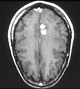
Behcet’s disease is most common along the “Old Silk Route,” which spans the region from Japan and China in the Far East to the Mediterranean Sea, including countries such as Turkey and Iran. Although the disease is rare in the United States, sporadic cases do occur in patients who would not appear to be at risk because of their ethnic backgrounds (e.g., in Caucasians or African–Americans). The disease is not rare in regions along the Old Silk Route, but the disease’s epidemiology is not well understood. In Japan, Behcet’s disease ranks as a leading cause of blindness. Below is a magnetic resonance image (MRI) study of a Behcet’s patient demonstrating central nervous system involvement (white matter changes in the pons).

Classic symptoms and signs of Behcet’s Disease

Behcet’s disease is virtually unparalleled among the vasculitides in its ability to involve blood vessels of nearly all sizes and types, ranging from small arteries to large ones, and involving veins too. Because of the diversity of blood vessels it affects, manifestations of Behcet’s may occur at many sites throughout the body. However, the disease has a predilection for certain organs and tissues; these are described below.
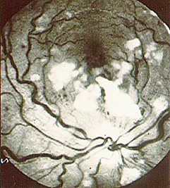
  • Eye
  • Mouth
  • Skin
  • Lungs
  • Joints
  • Brain
  • Genitals
  • Gastrointestinal Tract

Eye

  • Behcet’s may cause either anterior uveitis (inflammation in the front of the eye) or posterior uveitis (inflammation in the back of the eye), and sometimes causes both at the same time.
  • Anterior uveitis results in pain, blurry vision, light sensitivity, tearing, or redness of the eye.
  • Posterior uveitis may be more dangerous and vision–threatening because it often causes fewer symptoms while damaging a crucial part of the eye — the retina.
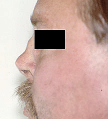
(top of section)

Mouth

  • Painful sores in the mouth called “aphthous ulcers”(pictured below). These are very similar in appearance to ulcers that frequently occur in the general population, usually as a result of minor trauma. In Behcet’s, however, the lesions are more numerous, more frequent, and often larger and more painful. Aphthous ulcers can be found on the lips, tongue, and inside of the cheek. Aphthous ulcers may occur singly or in clusters, but occur in virtually all patients with Behcet’s.

Skin

  • Pustular skin lesions that resemble acne, but can occur nearly anywhere on the body. This rash is sometimes called “folliculitis”.
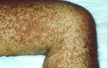
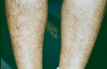
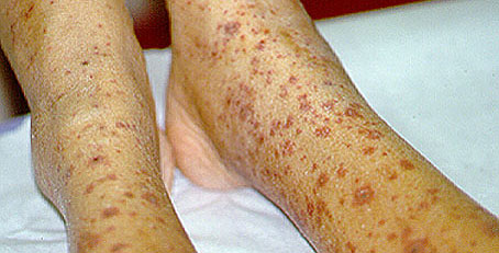
  • Skin lesions called erythema nodosum: red, tender nodules that usually occur on the legs and ankles but also appear sometimes on the face, neck, or arms. Unlike erythema nodosum associated with other diseases (which heal without scars), the lesions of Behcet’s disease frequently ulcerate.
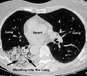
Lungs

  • Aneurysms (outpouchings of blood vessel walls, caused by inflammation) of arteries in the lung, rupture of which may lead to massive lung hemorrhage.

Joints

  • Arthritis or “arthralgias” (pain in the joints not accompanied by joint swelling).

Brain

  • Central nervous system involvement is one of the most dangerous manifestations of Behcet’s. The disease tends to involve the “white matter” portion of the brain and brainstem, and may lead to headaches, confusion, strokes, personality changes, and (rarely) dementia. Behcet’s may also involve the protective layers around the brain (the meninges), leading to meningitis. Because the meningitis of Behcet’s disease is not associated with any known infection, it is often referred to as “aseptic” meningitis.

Genitals

  • Male — painful genital lesions that form on the scrotum, similar to oral lesions, but deeper.
  • Female — painful genital ulcers that develop on the vulva.

Gastrointestinal

  • Ulcerations may occur anywhere in the gastrointestinal tract from the mouth to the anus. The terminal ileum and cecum are common sites. Involvement of the GI tract by Behcet’s may be difficult to distinguish from inflammatory bowel disease (such as Crohn’s disease).

Blood Vessels

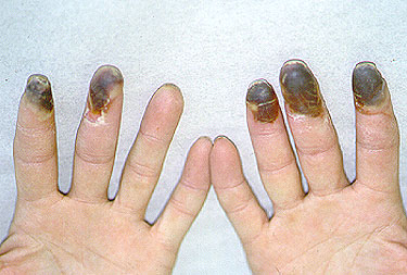
  • Clots can occur in veins in any site, most often including veins in the lower extremity (superficial or deep venous thrombosis).
  • Inflammation in arteries can occur as well, such as the pulmonary or abdominal arteries, sometimes causing obstruction of the vessel (thrombosis).

What causes Behcet’s Disease?

Behcet’s is one of the few forms of vasculitis in which there is a known genetic predisposition. The presence of the gene HLA–B51 is a risk factor for this disease. However, it must be emphasized that presence of the gene in and of itself is not enough to cause Behcet’s: many people possess the gene, but relatively few develop Behcet’s. Despite the predisposition to Behcet’s conferred by HLA–B51, familial cases are not the rule, constituting only about 5% of cases. Thus, it is believed that other factors (perhaps more than one) play a role. Possibilities include infections and other environmental exposures.

How is Behcet’s Disease Diagnosed?

There is not one specific test to diagnose Behcet’s. Rather the diagnosis is based on the occurrence of symptoms and signs that are compatible with the disease. The presence of certain features that are particularly characteristic (e.g., oral or genital ulcerations), elimination of other possible causes of the patient’s symptoms, and if possible, proof of vasculitis by biopsy of an involved organ would together support a diagnosis of Behcet’s.

A positive pathergy test can be supportive of the diagnosis of Behcet’s but is not diagnostic by itself of the condition. A pathergy test is a simple test in which the forearm is pricked with a small, sterile needle. Occurrence of a small red bump or pustule at the site of needle insertion constitutes a positive test. Please note, that although a positive pathergy test is helpful in the diagnosis of Behcet’s, only a minority of Behcet’s patients demonstrate the pathergy phenomenon (i.e., have positive tests). Patients from the Mediterranean region are more likely to demonstrate pathergy. In addition, other conditions can occasionally result in positive pathergy tests, so the test is not 100% specific.

Pictured below is an example of the pathergy test; 1) taken at the time when the patient was “stuck” with the sterile needle; 2) shows the area immediately after the stick; 3) & 4) show the area one day and two days after the needle stick, respectively.

Treatment and Course of Behcet’s Disease

For disease that is confined to mucocutaneous regions (mouth, genitals, and skin), topical steroids and non–immunosuppressive medications such as colchicine or dapsone may be effective. Apremilast (Otezla) is now FDA-approved for treatment of oral ulcers in Behcet’s. Moderate doses of systemic corticosteroids are also frequently required for disease exacerbations. Some patients require chronic, low doses of prednisone or conventional immunosuppressives such as (azathioprine) to keep the disease under control.

In the event of serious end–organ involvement such as eye or central nervous system disease, both high doses of prednisone and some other form of immunosuppressive treatment are usually necessary. Immunosuppressive agents used in the treatment of Behcet’s include azathioprine, cyclosporine, cyclophosphamide, and TNF-alpha inhibitors (infliximab, adalimumuab). Cyclophosphamide is generally used in life-threatening disease, such as central nervous system involvement. Blood clots can be another manifestation of Behcet’s, and in some scenarios blood thinners may be used in treatment.


Meet Our Team

Vasculitis Center Doctors

Duvuru Geetha, MD

Professor of Clinical Medicine

Dr. Geetha is a Professor of Medicine in the Division of Nephrology. A graduate of Madras Medical College, India, she completed Internal Medicine training in U.K. She did her Internal Medicine Residency at York, PA and Nephrology fellowship at Johns Hopkins Bayview Medical Center. She has been on Hopkins faculty since 1998. She is a member of Royal College of Physicians (U.K.), American Society of Nephrology, American Society of Transplantation and a consultant for the vasculitis foundation. She is a member of the Miller Coulson Academy of Clinical Excellence at Hopkins. Her clinical interests include renal disease in vasculitis patients with a focus on ANCA associated vasculitis and Henoch-Schonlein Purpura. She does clinical and translational research in vasculitis with a focus on ANCA associated vasculitis and renal disease.

Brendan Antiochos, MD

Assistant Professor of Medicine

Dr. Antiochos is a graduate of Dartmouth College and Dartmouth Medical School. He completed internal medicine residency at Oregon Health & Science University, then rheumatology fellowship at Johns Hopkins, before joining the faculty here in 2014. Dr. Antiochos assumed the role of Director for the vasculitis center in 2022. In addition to seeing patients in the vasculitis center, Dr. Antiochos performs laboratory-based research on autoimmune diseases. His laboratory work focuses on activation of the innate immune system and the discovery of novel autoantibodies.

Philip Seo, MD

Associate Professor of Medicine

A graduate of Harvard College and the College of Physicians and Surgeons at Columbia University, Dr. Seo completed his Internal Medicine training as a member of the Osler Medical Service at the Johns Hopkins Hospital. Since then, he has worked at Johns Hopkins in several capacities, including as a hospitalist at Johns Hopkins Bayview Medical Center, and as an Assistant Chief of Service of the Department of Medicine at the Johns Hopkins Hospital, before joining the Division of Rheumatology. His research interests are the assessment and treatment of ANCA-associated vasculitides, including Churg Strauss Syndrome, Wegener’s Granulomatosis, and Microscopic Polyangiitis.

David B. Hellmann, MD

Aliki Perroti Professor of Medicine

Dr. Hellmann is the Chairman of the Department of Medicine and Vice Dean at The Johns Hopkins Bayview Medical Center, and the Aliki Perroti Professor of Medicine. A graduate of Yale University and Johns Hopkins Medical School, Dr. Hellmann received his Internal Medicine training on the Osler Service at Hopkins, and trained in Rheumatology at the University of California, San Francisco. He has been on the Johns Hopkins faculty since 1986.

Desh Nepal, MD

Assistant Professor of Medicine

Michael Cammarata, MD

Assistant Professor of Medicine

Dr. Cammarata is a graduate of The College of William & Mary. He attended Eastern Virginia Medical School and completed his residency in Internal Medicine at the University of California San Francisco. He returned to the east coast for rheumatology fellowship at Johns Hopkins, joining faculty in 2024. He is RhMSUS certified in musculoskeletal ultrasound, and also practices general medicine as a hospitalist at Johns Hopkins Hospital. 

Collaborators

Vasculitis can involve virtually any organ system within the body. Hence, our Vasculitis Center maintains close collaborative relationships with experts from other specialties. The Vasculitis Center includes collaborators from several medical disciplines who help provide the highest level of care for our patients. They have extensive experience managing vasculitis within their subspecialty and work closely with the Physicians in the Vasculitis Center to provide comprehensive care for our patients:

Otolaryngology (ENT):

Our ENT team includes specialists in inflammatory sinus disease, sensorineural hearing loss, and chronic middle ear disease. We are pleased to also have a Doctor of Audiology, Dr. Dinkes, who specializes in inflammatory process on our team as well.

  • Dr. Jean Kim (sinus disease, middle ear manifestations)
  • Dr. Alexander Hiller (upper airway disease)
  • Dr. Roni Dinkes (audiology)

Neuro-ophthalmology:

  • Dr. Andrew Carey

Endocrinology / Osteoporosis:

  • Dr. Han Na Kim

Buerger’s Disease

  • First Description
  • Who gets Buerger’s Disease (the “typical” patients)?
  • Classic symptoms of Buerger’s Disease
  • What causes Buerger’s Disease?
  • How is Buerger’s Disease diagnosed?
  • Treatment and Course of Buerger’s Disease

First Description

This disease was first reported by Buerger in 1908, who described a disease in which the characteristic pathologic findings — acute inflammation and thrombosis (clotting) of arteries and veins — affected the hands and feet. Another name for Buerger’s Disease is thromboangiitis obliterans.

Who gets Buerger’s Disease (the “typical” patient)?

The classic Buerger’s Disease patient is a young male (e.g., 20–40 years old) who is a heavy cigarette smoker. More recently, however, a higher percentage of women and people over the age of 50 have been recognized to have this disease. Buerger’s disease is most common in the Orient, Southeast Asia, India and the Middle East, but appears to be rare among African–Americans.

Classic symptoms and signs of Buerger’s Disease

The initial symptoms of Buerger’s Disease often include claudication (pain induced by insufficient blood flow during exercise) in the feet and/or hands, or pain in these areas at rest. The pain typically begins in the extremities but may radiate to other (more central) parts of the body. Other signs and symptoms of this disease may include numbness and/or tingling in the limbs and Raynaud’s phenomenon (a condition in which the distal extremities — fingers, toes, hands, feet — turn white upon exposure to cold). Skin ulcerations and gangrene (pictured below) of the digits (fingers and toes) are common in Buerger’s disease. Pain may be very intense in the affected regions.

An angiogram demonstrating lack of blood flow to vessels of the hand (figure below). This decreased blood flow (“ischemia”) led to ulcers of the fingers and severe pain.

An abnormal result from an angiogram of the hand (figure below).

Despite the severity of ischemia (lack of blood flow) to the distal extremities that occurs in Buerger’s, the disease does not involve other organs, unlike many other forms of vasculitis. Even as ulcers and gangrene develop in the digits, organs such as the lung, kidneys, brain, and gastrointestinal (GI) tract remain unaffected. The reasons for the confinement to the extremities and sparing of other organs are not known.

What Causes Buerger’s Disease?

The association of Buerger’s Disease with tobacco use, particularly cigarette smoking, cannot be overemphasized. Most patients with Buerger’s are heavy smokers, but some cases occur in patients who smoke “moderately”; others have been reported in users of smokeless tobacco. It has been postulated that Buerger’s Disease is an “autoimmune” reaction (one in which the body’s immune system attacks the body’s own tissues) triggered by some constituent of tobacco.

Pictured below, are a patient’s fingertips that have developed gangrene. This is a very painful condition which sometimes requires amputation of the affected area.

How is Buerger’s diagnosed?

Buerger’s disease can be mimicked by a wide variety of other diseases that cause diminished blood flow to the extremities. These other disorders must be ruled out with an aggressive evaluation, because their treatments differ substantially from that of Buerger’s Disease (for Buerger’s, there is only one treatment known to be effective: complete smoking cessation — see below).

Diseases with which Buerger’s Disease may be confused include atherosclerosis (build–up of cholesterol plaques in the arteries), endocarditis (an infection of the lining of the heart), other types of vasculitis, severe Raynaud’s phenomenon associated with connective tissue disorders (e.g., lupus or scleroderma), clotting disorders of the blood, and others.

It should be noted that other substances, such as marijuana, have also been associated with a vasculitis similar to Buerger’s or polyarteritis nodosa that should be considered in the differential diagnosis.

Angiograms of the upper and lower extremities can be helpful in making the diagnosis of Buerger’s disease. In the proper clinical setting, certain angiographic findings are diagnostic of Buerger’s. These findings include a “corkscrew” appearance of arteries that result from vascular damage, particularly the arteries in the region of the wrists and ankles. Angiograms may also show occlusions (blockages) or stenoses (narrowings) in multiple areas of both the arms and legs.

Pictured below on the left is a normal angiogram. On the right, is an abnormal angiogram of an arm demonstrating the classic “corkscrew” appearance of arteries to the hand. The changes are particularly apparent in the blood vessels in the lower right hand portion of the picture (the ulnar artery distribution).

In order to rule out other forms of vasculitis (by excluding involvement of vascular regions atypical for Buerger’s), it is sometimes necessary to perform angiograms of other body regions (e.g., a mesenteric angiogram).

Skin biopsies of affected extremities are rarely performed because of the frequent concern that a biopsy site near an area poorly perfused with blood will not heal well.

Treatment and Course of Buerger’s

It is essential that patients with Buerger’s disease stop smoking immediately and completely. This is the only treatment known to be effective in Buerger’s disease. Patients who continue to smoke are generally the ones who require amputation of fingers and toes.

Despite the clear presence of inflammation in this disorder, anti-inflammatory agents such as steroids have not been shown to be beneficial. Similarly, strategies of anticoagulation (thinning of the blood with aspirin or other agents to prevent clots) have not proven effective. The only way to prevent the progression of the disease is to abstain from all tobacco products.

Linda’s Loop

contributed by Brenda Shilling

I have always known that chronic and life-threatening diseases can have a devastating effect on people’s lives. I’ve seen it happen to friends’ families and heard of it happening to friends of friends. But until 2002, the realities of such challenges were quite remote to me, as I had never felt a loved one struggle with a serious condition.

Linda Gray and her twin sister, Brenda Shilling.

In the spring of 2002, my identical twin, Linda, was diagnosed with neuropsychiatry lupus and central nervous system vasculitis. It was a scary time for all of us, particularly for Linda and her family. Like many lupus patients, Linda had been ill often and had many problems in the years leading up to her diagnosis. We were extremely thankful that The Johns Hopkins Vasculitis Center quickly identified what was wrong and started Linda on a treatment program.

In the months that followed, I watched my sister experience a multitude of emotions, including devastation over her diagnosis, fear for her future, and relief that she had finally found help. Although I know that not all lupus patients fare well, I learned that the treatment of lupus has come a long way over the years and I prayed that Linda would benefit from these advances. Her treatment was extremely challenging. However, during the hard realities of chemotherapy and steroids on Linda’s body, her spirit was amazing.

Despite knowing that family is extremely important during times like this and that frequent calls, letters, and visits to Linda were supportive, I slowly began to feel helpless. This was an unexpected emotion. I wanted to do more. I needed to do more. My sister was going through the most difficult time of her life, and I had do something more.

One morning in the late spring of 2003 I considered coordinating a bicycle ride to raise funds for The Johns Hopkins Vasculitis Center. Our sister, Liz, thought it was wonderful idea and wanted to help. We were driven by love for our sister and that was all the motivation we needed!

Linda Gray and her sister, Liz Adams.

We decided on a 50-mile ride – short enough to manage but long enough to sound good! We then mailed over 200 letters to friends and family asking for a financial gift to The Johns Hopkins Vasculitis Center in honor of our sister and others with her disease. We also distributed the letter to social and religious groups in which we are involved. Four of our friends asked if they could join us on the bike ride in a generous gesture of support. We even had t-shirts made!

Linda's Loop Participants

Participants in Linda’s Loop:

Left to Right: (Front Row, kneeling) Georgann Pattillo, Jan Rowe, Bill Schilling ; (Middle Row) Kevin Adams, Linda Moore, Liz Adams, Sue Schilling, Brenda Schilling, Betty Lamey, Patrick Pattillo, Leroy Lamey ; (Back Row) Chandler Burroughs, Steven Rowe, and Bill Schilling, Sr.

The night before the ride, we finished packing up drinks, snacks, and lunches and then headed home to get some sleep. Liz told me that she didn’t sleep a wink that night – neither did I! We were too excited.

The day finally arrived and everything went so well. It was such a wonderful day that I simply didn’t want it to end! Our family and friends came out to cheer us along. It felt great to have them there. But wow! The miles were more difficult than I expected. Although three riders finished far ahead, the remaining three of us dragged ourselves over the finish line some time later. We were quite the motley crew!

Hot, sweaty, hungry, but happy!

Response to our effort was overwhelming. My husband Bill teases that this was the only time I actually picked up the mail! Liz and I were moved by all the contributions. Some were from people who don’t know Linda but had read about the ride in the local paper. When we started planning we weren’t sure how much money we could raise. It was wonderful to have received just over $8,600 in contributions to vasculitis research!

Linda told me how much love she felt because of what we did for The Johns Hopkins Vasculitis Center. What more could I have asked of myself? We wanted to show Linda how proud we were of all she accomplished during her difficult treatment. We also wanted to say thank you to The Johns Hopkins Vasculitis Center for the wonderful work they do in caring for their patients everyday.

Hopefully we accomplished both.

All information contained within the Johns Hopkins Vasculitis Center website is intended for educational purposes only. Visitors are encouraged to consult other sources and confirm the information contained within this site. Consumers should never disregard medical advice or delay in seeking it because of something they may have read on this website.

Symptoms of Vasculitis

The term “vasculitis” refers to a group of inflammatory diseases that cause inflammation centered in the wall of blood vessels. This vascular inflammation ultimately leads to damage and dysfunction of the organs that contain the affected vessels. The symptoms of vasculitis depend on the particular blood vessels (and organs) that are involved by the inflammatory process.

As a group, the vasculitis syndromes have the ability to affect nearly every organ in the body. Yet different forms of vasculitis tend to involve blood vessels in specific locations throughout the body. For example, Giant Cell Arteritis typically involves the medium– to large–sized blood vessels supplying the head and neck, but rarely involves the blood vessels of the kidneys. In contrast, Granulomatosis with Polyangiitis (GPA) frequently involves the kidneys, very often the lungs, and almost always the upper respiratory tract, but rarely blood vessels to the brain.

buergerslg

As depicted in the image, Buerger’s disease involves the fingers (and toes). Gangrene can result from a profound lack of blood flow to these affected tissues.

Different types of vasculitis have characteristic (localized) patterns of blood vessel involvement.  However, vasculitis is often a systemic illness. Thus, patients with vasculitis generally feel sick. They often have fevers, weight loss, fatigue, a rapid pulse, and diffuse aches and pains that are difficult to pinpoint. It has been said that vasculitis is a “hurting disease”, because it is so commonly associated with pain of one type or another: pain from a nerve infarction, pain from insufficient blood to the gastrointestinal tract, pain from skin ulcers. In some cases, however, identifying the source and underlying cause of the pain is extremely challenging. Because vasculitis can involve virtually every organ system in the body, it often masquerades as other diseases, and may be a challenging diagnosis to make.

What organ systems may be affected?

It is important to note that not every organ system will be affected in every patient. The pattern of organ involvement (and symptoms) is unique to the individual, as well as the type of vasculitis (category).

Skin

A variety of rashes, the most classic of which is “palpable purpura” –purplish–red spots, usually found on the legs. These spots can usually be felt by the examiner’s fingertips, hence the descriptor “palpable”.

Purpura

This is a classic example of palpable purpura. These lesions result from the leakage of blood into the skin through inflamed, damaged blood vessels. They tend to occur in “crops”. This type of vasculitis involves very small diameter blood vessels in the skin.

Skin

Repeated bouts of purpura may lead to hyperpigmented (darkened) areas of the skin.

Joints

Symptoms range from full–blown arthritis to aches in the joints without obvious swelling (arthralgias).

Leurpura

This is an example of Henoch-Schönlein purpura: cutaneous vasculitis manifested by palpable purpura and arthritis (note the right ankle swelling). The diagnosis was confirmed by a skin biopsy, with immunofluorescence positive for IgA deposition witin blood vessel walls.

Lungs

Cough (particularly coughing up blood), shortness of breath, a pneumonia–like appearance to a patient’s chest X–ray, lung “infiltrates”, and the development of cavities in the lungs are among the manifestations that may occur in forms of vasculitis with lung involvement.

Lungs

This image comes from a CAT scan of the lungs of a 73 year–old woman complaining of constitutional symptoms, shortness of breath, and bloody sputum. The patient also had glomerulonephritis (kidney inflammation), a positive P–ANCA antibody test, and antibodies to myeloperoxidase. The diagnosis of microscopic polyangiitis was made.

Chest Xray

Eleven days later, as the patient’s symptoms worsened, a chest X–ray confirmed progression of her lung hemorrhage. The X–ray shows fluffy infiltrates in both lungs, representing bleeding from damaged capillaries.

Kidneys

In contrast to many other organs, inflammation in the kidneys does not hurt or cause other symptoms a patient would notice until renal damage is quite advanced. Instead, evidence of vasculitis involving the kidneys is made by obtaining lab tests, and in many cases a kidney biopsy. Glomerulonephritis is the most common type of kidney damage encountered in vasculitis. This syndrome can cause abnormal lab findings in the urine, including the presence of red blood cells (usually invisible to the naked eye), clumps of red blood cells (known as “casts”, also invisible to the naked eye), and abnormal levels of protein in the urine. If renal involvement is not recognized, renal failure can develop, sometimes leading to the need for dialysis or kidney transplant.

Glomerulus

Depicted in the figure to the right is a single glomerulus (the filtering unit of the kidneys; each kidney has approximately 1 million glomeruli) as seen on a kidney biopsy. The glomerulus is the part of the kidney that is affected by small vessel vasculitis, such as ANCA associated vasculitis.

angiogram

Polyarteritis nodosa (PAN) is another form of vasculitis that can involve the kidneys. PAN involves larger arteries that supply blood to the kidneys, and can be diagnosed using an angiogram (shown at right).

Blood

Vasculitis can cause abnormal findings on blood counts. Anemia (low hematocrit or red blood cell count) is a typical finding in patients with active vasculitis. A slightly elevated white blood cell count may also occur. These findings are very non-specific, meaning that they can occur in many other situations and diseases. Elevated inflammatory markers (erythrocyte sedimentation rate and C-reactive protein) are also frequently identified in patients with active vasculitis.

Sinus, Nose & Ears

The sinuses, nose and ears are common sites of involvement by ANCA-associated vasculitis, including GPA, EGPA and MPA. Symptoms can include chronic sinus congestion and “infections” that persist for longer than they should and require repeated courses of antibiotics; bleeding from the nose; perforations (holes) in the nasal septum; hearing loss; inflammatory fluid in the ears requiring drainage; inflammation in the cartilage of the ears or nose.

Nose

GPA involvement in the nose can lead to collapse of the nasal bridge (“saddle nose deformity”), as shown in the picture to the right.

Eyes

arterieslg

Vasculitis involving the eye may affect either blood vessels to the eyes, causing the sudden loss of vision, or small blood vessels within the eyes, leading to retinal problems, thinning of the sclera (the white part of the eyes), inflammation within the eye’s different chambers, and conjunctivitis (“pinkeye”). Pictured at right is an example of retinal vasculitis in a patient with systemic lupus erythematosus (lupus). The white areas represent regions of retinal infarction caused by vasculitis. The most feared complication of GCA is blindness caused by injury to the optic nerve.

Brain

brainhemlg

The brain is not a common site of vasculitis involvement. When present, vasculitis in this site can cause headaches, strokes, changes in mental status, or difficulty with coordination. At right, a magnetic resonance (MR) imaging study of the brain in central nervous system vasculitis demonstrates an intra–cerebral hemorrhage (bright area).

Nerve

Peripheral nerves are a relatively common site of vasculitis involvement. Damage to the peripheral nerves can cause shooting pains in the arms and legs, numbness, and asymmetrical weakness (i.e., weakness that involves one side of the body more than the other).

All information contained within the Johns Hopkins Vasculitis Center website is intended for educational purposes only. Visitors are encouraged to consult other sources and confirm the information contained within this site. Consumers should never disregard medical advice or delay in seeking it because of something they may have read on this website.

Eosinophilic Granulomatosis with Polyangiitis, formerly Churg-Strauss Syndrome (EGPA)

  • First Description
  • Who gets EGPA (the “typical” patients)?
  • Classic symptoms of EGPA
  • What causes EGPA?
  • How is EGPA diagnosed?
  • Treatment and Course of EGPA

First Description

Eosinophilic granulomatosis with polyangiitis (EGP), formerly known as the Churg-Strauss Syndrome , is a systemic vasculitis. This disease was first described in 1951 by Dr. Jacob Churg and Dr. Lotte Strauss as a syndrome consisting of “asthma, eosinophilia [an excessive number of eosinophils in the blood], fever, and accompanying vasculitis of various organ systems”. EGPA shares many of the clinical and pathological features of polyarteritis nodosa (“PAN”, another type of vasculitis) and granulomatosis with polyangiitis (GPA). However, Drs. Churg and Strauss discovered that the presence of granulomas as well as the abundance of eosinophils distinguished this disease from PAN and GPA.

Who gets EGPA (the “typical” patient)?

The typical patient with EGPA is a middle aged individual with a history of new-onset or newly-worsened asthma. The distribution of the disease among males and females is approximately equal.

Classic symptoms and signs of EGPA

Asthma is one of the cardinal features of EGPA. Asthma symptoms may begin long before the onset of vasculitis – e.g., many years before any other symptoms of EGPA arise, and long before the diagnosis of EGPA is made. Other early symptoms/signs include nasal polyps and allergic rhinitis.

The next phase of the disease is often marked by eosinophilia, the finding of an excessive number of eosinophils in the blood or in tissues. An eosinophil is one subtypes of white blood cell. Normally, eosinophils comprise 5% or less of the total white blood cell count. In EGPA, the percentage of eosinophils may reach as high as 60%. In the picture below, the eosinophils are shown by the dark pink stain.

The third phase of the illness is a vasculitis, which involves the skin, lungs, nerves, kidneys, and other organs. Particular mention should be made of the frequent devastating involvement of the nerves (called mononeuritis multiplex), which produces severe tingling, numbess, shooting pains, and severe muscle wasting/power loss in the hands or feet. The list below contains the organs commonly involved by EGPA and the specific disease manifestation(s) in each organ.

  • Nose
  • Lung
  • Skin
  • Kidney
  • Gastrointestinal
  • Heart
  • Nerve

Nose

  • Sinusitis, including allergic rhinitis
  • Nasal polyps

Lung

  • Pulmonary infiltrates (only one-third of all patients)
  • Bleeding into the lungs (occasionally)
  • Diffuse interstitial lung disease (rarely)

Skin

  • Rashes
  • Palpable purpura
  • Nodules (above or below the skin), often at sites of pressure, such as the elbows

Kidney

  • Glomerulonephritis (inflammation in the small units of the kidney that filter blood)
  • Hypertension

Gastrointestinal

  • Lesions (vasculitic) are occasionally found in the GI tract
  • Granuloma sometimes found in spleen

Heart

  • Vasculitis lesions in heart, can lead to congestive heart failure or a heart attack

Nerve

  • Peripheral nerve involvement including pain, numbness, or tingling in extremities (neuropathy/mononeuritis multiplex)

What causes EGPA?

The cause of EGPA is unknown but is probably multi-factorial. Genetics may play a small role in the disease, but EGPA is almost never seen in two members of the same family. Environmental factors such as exposure to industrial solvents may play a role in susceptibility to this disease, but this is largely speculative. Infections may be the inciting event(s), but to date there is no definitive evidence of this.

How is EGPA Diagnosed?

Among all of the vasculitides, asthma is a distinctive feature of EGPA alone. However, not all patients with asthma have vasculitis – only a tiny minority do, in fact. It is the specific combination of symptoms and signs, the pattern of organ involvement, and the presence of certain abnormal blood tests (eosinophilia, in particular) that help the doctor make the diagnosis. In addition to a detailed history and physical examination, blood tests, chest X-rays and other types of imaging studies, nerve conduction tests, and tissue biopsies (e.g., of lung, skin, or nerve) may be performed to help diagnose EGPA.

The following features are consistent with a diagnosis of GPA:

  1. asthma
  2. eosinophilia [>10% on differential WBC count]
  3. mononeuropathy
  4. transient pulmonary infiltrates on chest X-rays
  5. paranasal sinus abnormalities
  6. biopsy containing a blood vessel with extravascular eosinophils.

Treatment and Course of EGPA

EGPA usually responds to prednisone. Initially, high doses of oral prednisone are used in an attempt to get the disease into remission as quickly as possibly (e.g., using oral prednisone 40-60 mg/day). After the first month or so, this high dose of prednisone is gradually tapered down over the ensuing months. Other immunosuppressive drugs, such as azathioprine, cellcept, methotrexate, cyclophosphamide, or rituximab may be used in addition to prednisone. High doses of intravenous steroids (usually methylprednisolone) maybe useful for those patients with severe disease or for those who are unresponsive to the combination of oral prednisone used with other immunosuppressive medications.

Prior to the advent of prednisone, EGPA was often a fatal disease. The majority of patients died from rampant, uncontrolled disease. With present therapy, constitutional symptoms begin to resolve quite quickly, with gradual improvement in cardiac and renal function, as well as improvement in the pain that results from peripheral nerve involvement. The course of therapy can last for 1 to 2 years, although the length and type of treatment depend on the severity of disease and the organs involved. The patient’s response to treatment and the continuation of disease control during lowering of the prednisone dose are the primary determinants of how long therapy is continued. Laboratory monitoring of blood tests is very helpful in gauging the activity of disease. Some of the most useful laboratory tests are the erythrocyte sedimentation rate (ESR) and the eosinophil count.

  • « Go to Previous Page
  • Go to page 1
  • Go to page 2
  • Go to page 3
  • Go to page 4
  • Go to page 5
  • Go to Next Page »

Primary Sidebar


donate to the Johns Hopkins Vasculitis Center

All information contained within the Johns Hopkins Vasculitis Center website is intended for educational purposes only. Visitors are encouraged to consult other sources and confirm the information contained within this site. Consumers should never disregard medical advice or delay in seeking it because of something they may have read on this website.

Footer

Johns Hopkins Rheumatology

  • Johns Hopkins Rheumatology
  • Johns Hopkins Arthritis Center
  • Johns Hopkins Lupus Center
  • Johns Hopkins Lyme Disease Research Center
  • Johns Hopkins Myositis Center
  • Johns Hopkins Scleroderma Center
  • Johns Hopkins Sjögren’s Syndrome Center

Connect With Us

  • Facebook
  • Twitter
  • YouTube
U.S. News and World Report Rankings Badge

Johns Hopkins Medicine Johns Hopkins Medicine logo
Language Assistance Available:
  • Español
  • American Sign Language
  • עִברִי
  • אידיש
  • አማርኛ
  • 繁體中文
  • Français
  • Tagalog
  • Русский
  • Português
  • Italiano
  • Tiếng Việt
  • Ɓàsɔ́ɔ̀-wùɖù-po-nyɔ̀
  • Igbo asusu
  • èdè Yorùbá
  • বাংলা
  • 日本語
  • 한국어
  • Kreyòl Ayisyen
  • العربية
  • Deutsch
  • Polski
  • Ελληνικά
  • ગુજરાતી
  • ภาษาไทย
  • اُردُو
  • فارسی
  • हिंदी
  • Deitsch
  • ខ្មែរ
  • မြန်မာ
Contact & Privacy Information
Price Transparency
Notice of Privacy Practices Privacy Statement
Terms & Conditions of Use Non-Discrimination Notice
Manage Cookie Preferences
Copyright © 2025 Johns Hopkins Rheumatology