- Who gets Granulomatosis with Polyangiitis (the “typical” patients)?
- Classic symptoms of Granulomatosis with Polyangiitis
- What causes Granulomatosis with Polyangiitis?
- How is Granulomatosis with Polyangiitis diagnosed?
- Treatment and Course of Granulomatosis with Polyangiitis
Who gets Granulomatosis with Polyangiitis?
Granulomatosis with Polyangiitis is nearly equally distributed between the sexes, with a slight male predominance. Granulomatosis with Polyangiitis typically occurs in middle age, but is found in people of all ages. Although it is unusual for Granulomatosis with Polyangiitis to occur in childhood, it is not unusual for a Granulomatosis with Polyangiitis patient to be in his/her 70s or even 80s at the time of diagnosis.
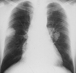
Pictured below is a chest x–ray showing bilateral lung nodules in a 27 year old Indian man with Granulomatosis with Polyangiitis.
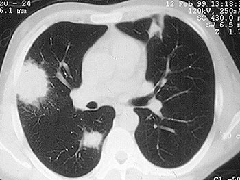
Pictured below is a CT scan from the same patient. The view is a cross–section through the patient’s lungs. The CT scan not only permits a better appreciation of the lesions’ size, it also detects more lesions.
Granulomatosis with Polyangiitis can affect virtually any site in the body, but it has a predisposition for certain organs. The classic organs involved in Granulomatosis with Polyangiitis are the upper respiratory tract (sinuses, nose, ears, and trachea [the “windpipe”]), the lungs, and the kidneys. Listed below are the organs commonly involved in Granulomatosis with Polyangiitis and the specific disease manifestation(s) in each organ.
Ear
Inflammation in the ear can look and behave similarly to an ear infection, but recurs frequently and is not caused by infection. Patients may need to have tubes placed in the ears to drain inflammatory fluid. Hearing loss may result from damage to the middle ear. Inflammation in the inner ear can also cause hearing loss and balance disturbance (vertigo). The mastoid bone may be another site of GPA involvement (mastoiditis).
Eye
Inflammation can occur in different parts of the eye. Inflammation in the white part of the eye is known as the sclera (“scleritis”). “Uveitis” is inflammation within the eye. Inflammation behind the eye is known as an “orbital pseudotumor”. An orbital pseudotumor such as those caused by Granulomatosis with Polyangiitis can cause “proptosis”, or protrusion of one eye.
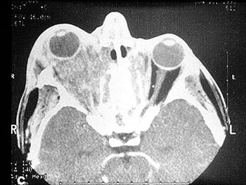
Pictured below is a computed tomography (CAT) scan of the eyes in a patient with a retro–orbital mass (a mass behind the eye) in a man with Granulomatosis with Polyangiitis. Masses such as these sometimes cause an abrupt loss of vision through stretching or compression of the optic nerve, which enters the back of the eye.
Nose
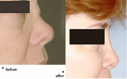
Nasal crusting and frequent nosebleeds can occur, along with erosion and perforation of the nasal septum. The bridge of the nose can collapse resulting in a “saddle–nose deformity”. Pictured below is an example of this deformity before and after cosmetic surgery. This resulted from the collapse of the nasal septum caused by cartilage inflammation. This patient has Granulomatosis with Polyangiitis, but an identical lesion may occur in Relapsing Polychondritis. This form of nasal collapse can occur relatively rapidly.
Sinuses
Chronic sinus inflammation that sometimes leads to a destructive process of tissues around the sinuses is nearly universal in GPA, and is often the site in which the disease begins. Most patients have been treated repeatedly for what is thought to be a sinus infection, but without success. Sinus surgeries are common among GPA patients.
Trachea
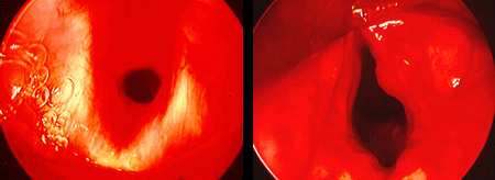
A characteristic respiratory tract complication of Granulomatosis with Polyangiitis: narrowing of the “windpipe” just below the vocal cords, a condition called “subglottic stenosis”. This narrowing, caused by inflammation and scarring, causes difficulty breathing and may, after a subacute progression, necessitate emergency tracheostomy. Pictured below are two figures that show subglottic stenosis before (left) and after (right) surgery, performed by an Ear, Nose, & Throat specialist. The surgery provided dramatic improvement in the patient’s breathing.
Lungs
GPA causes two main manifestations in the lungs. Lung nodules are mass lesions that look identical to infections or cancer on radiographic studies, but show evidence of inflammation on biopsy. These lung nodules may not cause any symptoms until seen on imaging studies. Bleeding into the lungs can also occur, in a syndrome known as “diffuse alveolar hemorrhage” – a potential emergency which is most often diagnosed and treated in the hospital.
Kidney
Inflammation can occur in the kidney, leading to small (or rarely, large) amounts of blood and protein in the urine. This condition is called glomerulonephritis. If not treated aggressively, Granulomatosis with Polyangiitis’s involvement of the kidneys can lead to kidney failure. Renal masses can occur, but are very unusual in this disease.
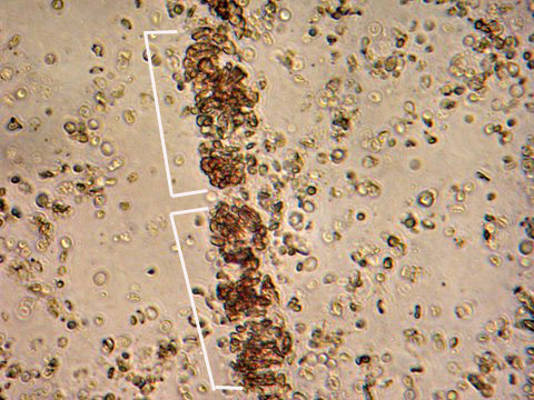
The image below is from a urinalysis of a patient with kidney inflammation. When Granulomatosis with Polyangiitis is active, red blood cells will form a clump or “cast” (bracketed in white) within the tubules of inflamed kidneys. These “casts” pass through the renal system and may be viewed under the microscope in a patient’s urine.
Skin
Granulomatosis with Polyangiitis can cause many kinds of skin rashes. The most common rash occurs in the form of small purple or red dots on the lower extremities (known as “palpable purpura”). Inadequate blood flow to fingers and toes can lead to Raynaud’s phenomenon (extreme sensitivity of the fingers to cold) and even infarctions of the tips of fingers and toes, with the development of gangrene.
Joints
Inflammation in the joints (arthritis) can occur, causing joint swelling, pain, and a sensation of stiffness. This sensation is typically worse in the mornings and improves with activity.
Nerves
Peripheral nerve involvement leads to numbness, tingling, shooting pains in the extremities, and sometimes to weakness in a foot, hand, arm, or leg. Painful weakness in the extremities represents a type of nerve inflammation called “mononeuritis multiplex” and is a medical emergency that requires prompt diagnosis and treatment.
Miscellaneous
Granulomatosis with Polyangiitis involvement of nearly all organs has been described, including the meninges (the layers of protective tissue around the brain and spinal cord), the prostate gland, and the genito–urinary tract. In addition to involving specific organs, Granulomatosis with Polyangiitis also commonly results in generalized symptoms of fatigue, low–grade fever, and weight loss.
The cause of Granulomatosis with Polyangiitis is not known. Compared to diseases with obvious genetic predispositions, genetics appear to play a relatively small role in the etiology of Granulomatosis with Polyangiitis . It is very unusual for Granulomatosis with Polyangiitis to occur in two people in the same family. (It is possible, however, that less obvious genetic risk factors exist, e.g. genes that might pre-–dispose a patient to infection with an etiologic organism). For some time, an infection has been suspected of causing (or at least contributing to) Granulomatosis with Polyangiitis , but no specific infection (bacterial, viral, fungal, or other) has been identified.
How is Granulomatosis with Polyangiitis Diagnosed?
Whenever possible, it is important to confirm the diagnosis of Granulomatosis with Polyangiitis by biopsying an involved organ and finding the pathologic features of this disease under the microscope. Because many diseases may mimic Granulomatosis with Polyangiitis (and vice versa), before starting a treatment regimen it is essential to be as certain of the diagnosis as possible. We discuss some of the specific biopsy procedures used to diagnose Granulomatosis with Polyangiitis in the section of this Websie entitled What is Vasculitis: Diagnosis?.
Because Granulomatosis with Polyangiitis so often involves the upper respiratory tract (sinuses, nose, ears, and trachea [“windpipe”]) and because biopsy of these tissues is a relatively non–invasive procedure, these sites are frequently biopsied in patients suspected of Granulomatosis with Polyangiitis . Unfortunately, the yield of biopsies from these sites is rather low: probably less than 50%. Therefore, sometimes more invasive procedures are required to make the diagnosis.
Lung biopsy (either open or thoracoscopic) is often the best way of diagnosing Granulomatosis with Polyangiitis . The ample amount of tissue obtainable through these procedures usually permits confirmation of the Granulomatosis with Polyangiitis diagnosis. Similarly, although the amount of tissue obtained through a kidney biopsy is usually much smaller, the finding of certain pathologic features in the context of a patient’s overall symptoms, signs, and laboratory tests is frequently diagnostic.
Since 1982, when ANCAs (anti–neutrophil cytoplasmic antibodies) were first described, the role of these antibodies in the diagnosis of Granulomatosis with Polyangiitis has grown. ANCA testing, which involves the performance of a simple blood test, has achieved wide availability during the 1990s. This is both good and bad: use of ANCA tests has led to earlier diagnoses and more rapid institution of appropriate treatment in many cases, but has also resulted in misdiagnosis and incorrect treatment when the tests are not performed or interpreted correctly.
As their name implies, ANCAs are directed against the cytoplasm (the non-nucleus part) of white blood cells. Their precise role in the disease process remains uncertain but is a topic of considerable research interest. ANCAs come in two primary forms: 1) the C–ANCA [C stands for cytoplasmic] and, 2) the P–ANCA [P stands for perinuclear]. C–ANCAs have a particularly strong connection to Granulomatosis with Polyangiitis (up to 80% of patients — and possibly more of those with active disease — have these antibodies). When C–ANCAs are present in the blood of a patient whose symptoms or signs suggest Granulomatosis with Polyangiitis , the likelihood of the diagnosis increases considerably. In most cases, however, it is still VERY IMPORTANT to biopsy an involved organ to verify the diagnosis.
Treatment and Course of Granulomatosis with Polyangiitis
Until the 1970s, Granulomatosis with Polyangiitis was nearly always a fatal condition. The use of prednisone and other steroids helped prolong patients’ lives, but most patients eventually succumbed to the disease within a few months or years. The first use of cyclophosphamide in the late 1960s began to change the terrible prognosis of this disease. Using the combination of cyclophosphamide and prednisone, more than 90% of patients with severe disease respond to treatment, and 75% are able to achieve disease remissions.
Today, Rituximab is the standard recommended therapy for patients with major organ involvement of GPA. The vast majority of patients respond very well to Rituximab, which is used both early in the disease course (induction treatment) and over longer timeframes (maintenance therapy).
Sometimes, patients with milder disease are treated with oral alternatives, such as methotrexate, leflunomide, or azathioprine.
Unfortunately, Granulomatosis with Polyangiitis is a disease in which relapses frequently occur. Approximately half of all patients who achieve disease remissions eventually suffer recurrences (“flares”). Flares of Granulomatosis with Polyangiitis are usually responsive to the same treatment that induced remission, but sometimes intensification of treatment (for example, changing to a more powerful medication) is required.”